Pericardial Cyst:
View cases of pericardial cysts
Clinical:
Pericardial cysts are benign lesions that account for 5-10% of all
mediastinal tumors [2]. True pericardial cysts are rare. They are
believed to be congenital abnormalities which
occur as the result of persistence of the ventral and parietal
pericardial recesses.
Pericardial cysts, however, do not communicate with the pericardial
space.
Pleuropericardial cysts are more common. Although present at birth, the
lesion is
asymptomatic and is usually not detected until adulthood. Pericardial
cysts occur most
commonly in the right cardiophrenic angle (60-70%), followed by the
left side (22-30%) [3].
About 8-10% of lesions occur in other sites- sometimes the cyst will be
pretracheal and
mimic a foregut cyst. Because of the benign nature of the lesion,
surgical resection should be performed only in patients with associated
symptoms [2]. Differential considerations for a typical pericardial
cyst include
an intrapericardial bronchogenic cyst, pericardial neoplasm, or
intrapericardial teratoma
(neonates- may appear cystic).A pericardial diverticulum is a focal
outpouching of the pericardial sac, which can be differentiated from a
pericardial cyst by the rpesence of a direct communication with the
pericardial cavity (usually identified by changes in size related to
body position) [3].
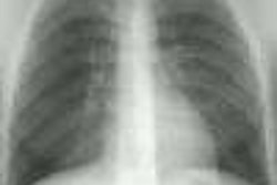
X-ray:
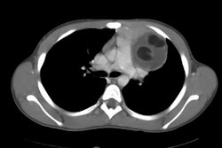
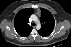
On CT the lesion appears as a smooth, sharply marginated, water density (HU 0-20) mass which typically touches the anterior chest wall, diaphragm, and heart. It is encapsulated by a very thin wall which may not be evident by CT. Mural calcification is rare. Higher attenuation values may occur due to prior intracyst hemorrhage or infection. On MRI, the lesion has low signal intensity on T1 and bright signal on T2 images.
REFERENCES:
(1) Chest 1997; Strollo DC, et al.
Primary
mediastinal
tumors. Part II: Tumors of the middle and posterior
mediastinum. 112: 1344-57
(2) Radiographics 2007; Pineda V, et al. Lesions of the
cardiophrenic space: findings at cross-sectional imaging. 27: 19-32
(3) Radiology 2013; Bogaert J, Francone M. Pericardial disease: value of CT and MR imaging. 267: 340-356