Malignant Tumor of Nerve Sheath Origin:
Clinical:
This tumor was formerly referred to as a malignant schwannoma or neurofibrosarcoma. The lesion is rare 5-10% of all soft tissue sarcomas, and intrathoracic MPNSTs are particularly rare [3] (the lesion is rare and accounts for less than 1% of all mediastinal neurogenic neoplasms). It is an aggressive, locally invasive, and metastasizing spindle cell sarcoma that typically arises from a simple or plexiform neurofibroma (and rarely, if ever, from a pre-existing schwannoma). Patients with neurofibromatosis (NF1) are at an increased risk for this tumor (about 20% to 50% of cases occur in patients with NF1 and the life time prevalence is about 10%), and have a significantly worse prognosis (5-year survival of approximately 34-60% [3]). Patients typically present around 40-50 years of age (NF1 patients present at a younger age [3]), with pain, a rapidly enlarging mass, or nerve deficit. The lesion frequently metastasizes hematogenously to the lungs.X-ray:
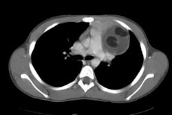
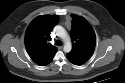
The lesion is typically large by the time of presentation. Low attenuation areas within the mass are related to the presence of hemorrhage and necrosis.On MR, findings which suggest a MPNST rather than a neurofibroma include a large lesion (>5cm), peripheral enhancement pattern, the presence of perilesional edma-like zone, and intratumoral cystic change [3]. The lesion is intensely FDG avid on PET imaging [3].
REFERENCES:
(1) Chest 1997; Strollo DC,
et
al. Primary mediastinal tumors. Part II: Tumors of the
middle and posterior
mediastinum. 112: 1344-57
(2) AJR 2007; Bredella MA, et al. Value of PET in the assessment
of patients with neurofibromatosis type I. 189: 928-935
(3) AJR 2016; Pavlus JD, et al. Imaging of thoracic neurogenic tumors. 207: 552-561