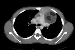
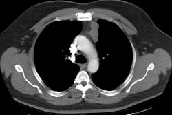
Myasthenia Gravis:
Myasthenia gravis is a T-cell dependent autoimmune disorder characterized by auto-antibodies that bind to acetylcholine receptors on the postjunctional muscle membrane and/or muscle specific tyrosine kinase autoantibodies, resulting in a progressive and occasionally generalized weakness of skeletal muscles. Although any muscle group can be involved, the muscles innervated by the cranial nerves (especially the extraoccular and facial muscles) are affected most frequently. Patients can present with ptosis, diplopia, dysphagia, dysarthria, drooling, and difficulty chewing. The most serious complication is myasthenic crisis which is characterized by life-threatening paralysis and/or respiratory failure- this can be seen in up to 27% of patients within 2 years of diagnosis [2].The most common associated thymic abnormality is follicular thymic hyperplasia (about 65% of cases), while thymoma occurs in about 10-15% of patients with the disease.
Iodinate contrast: The use of iodinated contrast, even low osmolar contrast, can precipitate an acute exacerbation of myasthenia gravis-related symptoms- most commonly progressive acute respiratory compromise which can result in death [2].
REFERENCES:
(1) Radiographics 2002; Santana L, et al. Best cases from the AFIP:
Thymoma.
22: S95-S102
(2) Radiology 2013; Somashekar DK, et al. Effect of intravenous
low-osmolality iodinated contrast media on patients with myasthenia
gravis. 267: 727-734