Lateral Thoracic Meningocele:
Clinical:
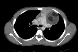
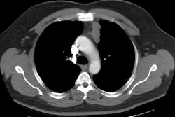
Represents a herniation of the leptomeninges through the neural foramina. It is a rare, typically asymptomatic lesion that occurs more commonly on the right side [2]. Bilateral meningoceles can be found in 10% of cases. Neurofibromatosis (Type 1) is seen in 75-85% of cases and sharp angle scoliosis is also frequently found in association with the lesion. Surgical ligation of the neck of the dural sac and resection of the meningocele is the usual treatment.X-ray:
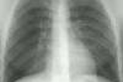
On CT the lesion appears as a homogeneous, water density paravertebral mass which may enlarge the neural foramina, cause posterior vertebral scalloping, or produce a scoliosis (found in 50% of cases- the meningocele is near the apex of the deformity on the convex side). The lesion does not enhance with contrast. Myelography can be used to demonstrate communication with the subarachnoid space. On MRI, the signal intensity of the fluid within the meningocele is identical to that of CSF.Lateral thoracic meningocele: The patient below presented with back pain. The CXR demonstrated a large posterior mediastinal mass. If you look carefully at the lateral view you can see that the neural foramina at the level of the lesion is expanded. CT scan demonstrated bilateral lateral thoracic meningoceles- larger on the right, with neural foramina expansion and posterior vertebral scalloping. MR demonstrates signal intensity identical to CSF. |
|


REFERENCES:
(1) Chest 1997; Strollo DC, et
al. Primary mediastinal tumors. Part II: Tumors of the middle and posterior
mediastinum. 112: 1344-57
(2) AJR 1999; Rossi SE, et al. Thoracic manifestations of Neurofibromatosis-I. 173: 1631-1638