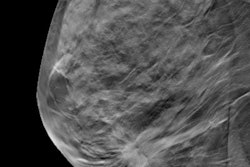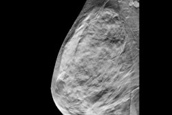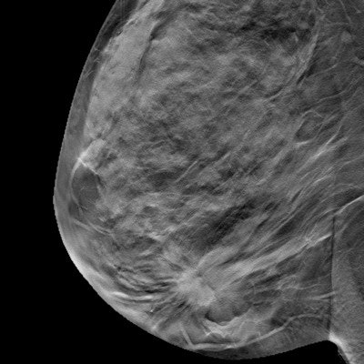
Imaging surveillance can be an alternative for managing architectural distortion on digital breast tomosynthesis (DBT) showing radial scarring without atypia, according to research published February 2 in the American Journal of Roentgenology.
Researchers led by Dr. Juan Villa Camacho from Massachusetts General Hospital n Boston found that such distortion on DBT with concordant nonmalignant pathology at biopsy has an overall upgrade rate to malignancy at surgery of 10.2%.
They also found that architectural distortion without atypia has a low upgrade rate of 2.2%, and 28.2% with atypia.
Surgery is the recommended course of action for all architectural distortion cases, including nonmalignant ones. However, with low upgrade rates seen in the study, the researchers wrote that surveillance can be employed.
The team looked at data from 129 cases of architectural distortion with nonmalignant pathology at biopsy that underwent excision in 125 women with an average age of 54 years.
Out of these, 92 were radial scars and 37 were other nonmalignant pathologies. The researchers reported that only one out of 66 radial scars without atypia at biopsy was upgraded to ductal carcinoma in situ (DCIS) at surgery, while none were upgraded to invasive cancer.
Of the 24 benign pathologies without atypia at biopsy found, the study authors reported that one was considered discordant. They also wrote that one of the remaining 23 concordant cases was upgraded to DCIS at surgery and none to invasive cancer.
Other features, such as age, personal or family breast cancer history, and breast density, among others showed no significant associations with upgrade risk.

![A normal mammogram confirmed by three-year radiologic follow-up illustrates reader-marked regions of interest (ROIs) during (A) unaided (round 1) and (B) artificial intelligence (AI)–assisted (round 2) reading. Each colored dot represents an ROI for recall by a human reader. Readers could mark more than one ROI per case, represented by multiple dots of the same color. During AI-assisted reading, the AI system displayed three visible prompts: two with suspicion of malignancy scores of 35% (left mediolateral oblique [L MLO] and craniocaudal [L CC]) and one with a suspicion of malignancy score of 10% (right craniocaudal [R CC]), shown as polygonal overlays. Without AI, six of 10 readers (60%) marked a false-positive ROI. With AI assistance, this fell to two of 10 (20%). R MLO = right mediolateral oblique.](https://img.auntminnie.com/mindful/smg/workspaces/default/uploads/2026/07/2026-07-14-radiology-mammogram-ai-auto-bias.H0bYO8QlWs.jpg?auto=format%2Ccompress&fit=crop&h=100&q=70&w=100)


![A normal mammogram confirmed by three-year radiologic follow-up illustrates reader-marked regions of interest (ROIs) during (A) unaided (round 1) and (B) artificial intelligence (AI)–assisted (round 2) reading. Each colored dot represents an ROI for recall by a human reader. Readers could mark more than one ROI per case, represented by multiple dots of the same color. During AI-assisted reading, the AI system displayed three visible prompts: two with suspicion of malignancy scores of 35% (left mediolateral oblique [L MLO] and craniocaudal [L CC]) and one with a suspicion of malignancy score of 10% (right craniocaudal [R CC]), shown as polygonal overlays. Without AI, six of 10 readers (60%) marked a false-positive ROI. With AI assistance, this fell to two of 10 (20%). R MLO = right mediolateral oblique.](https://img.auntminnie.com/mindful/smg/workspaces/default/uploads/2026/07/2026-07-14-radiology-mammogram-ai-auto-bias.H0bYO8QlWs.jpg?auto=format%2Ccompress&fit=crop&h=112&q=70&w=112)