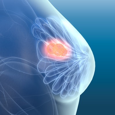
Microwave breast imaging may have the potential to be a noninvasive, nonionizing, and painless addition to the breast cancer diagnosis toolbox, according to research published August 4 in Academic Radiology.
In a "first-in-human" study, a team led by Brian Moloney, PhD, from Galway University Hospital in Ireland found that a microwave breast imaging system detected and localized the majority of breast lesions in 24 women.
Microwave breast imaging is an emerging technology that researchers have touted as a nonionizing, noncompressive approach to breast cancer diagnosis. However, current prototypes have been limited in localizing lesions.
For their study, Moloney's group used the Wavelia system developed by French firm MVG Industries; the device uses a low-power electromagnetic wave. Women were divided into three groups: those with biopsy-proven breast cancers, those with unaspirated cysts, and those with biopsy-proven benign breast lesions.
The system detected and localized 12 of 13 benign breast lesions, and nine out of the 11 breast cancers, including one case of a radiographically occult invasive lobular cancer. No device-related adverse events were recorded. Twenty-three of the women reported that they would recommend microwave breast imaging to other women.
While more studies need to be done with larger patient cohorts, the Wavelia system holds "significant" potential for detecting breast abnormalities while offering patients a favorable experience over conventional mammography, the authors concluded.

![A normal mammogram confirmed by three-year radiologic follow-up illustrates reader-marked regions of interest (ROIs) during (A) unaided (round 1) and (B) artificial intelligence (AI)–assisted (round 2) reading. Each colored dot represents an ROI for recall by a human reader. Readers could mark more than one ROI per case, represented by multiple dots of the same color. During AI-assisted reading, the AI system displayed three visible prompts: two with suspicion of malignancy scores of 35% (left mediolateral oblique [L MLO] and craniocaudal [L CC]) and one with a suspicion of malignancy score of 10% (right craniocaudal [R CC]), shown as polygonal overlays. Without AI, six of 10 readers (60%) marked a false-positive ROI. With AI assistance, this fell to two of 10 (20%). R MLO = right mediolateral oblique.](https://img.auntminnie.com/mindful/smg/workspaces/default/uploads/2026/07/2026-07-14-radiology-mammogram-ai-auto-bias.H0bYO8QlWs.jpg?auto=format%2Ccompress&fit=crop&h=100&q=70&w=100)


![A normal mammogram confirmed by three-year radiologic follow-up illustrates reader-marked regions of interest (ROIs) during (A) unaided (round 1) and (B) artificial intelligence (AI)–assisted (round 2) reading. Each colored dot represents an ROI for recall by a human reader. Readers could mark more than one ROI per case, represented by multiple dots of the same color. During AI-assisted reading, the AI system displayed three visible prompts: two with suspicion of malignancy scores of 35% (left mediolateral oblique [L MLO] and craniocaudal [L CC]) and one with a suspicion of malignancy score of 10% (right craniocaudal [R CC]), shown as polygonal overlays. Without AI, six of 10 readers (60%) marked a false-positive ROI. With AI assistance, this fell to two of 10 (20%). R MLO = right mediolateral oblique.](https://img.auntminnie.com/mindful/smg/workspaces/default/uploads/2026/07/2026-07-14-radiology-mammogram-ai-auto-bias.H0bYO8QlWs.jpg?auto=format%2Ccompress&fit=crop&h=112&q=70&w=112)