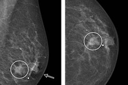
New research suggests removing additional tissue around the tumor cavity for patients with ductal carcinoma in situ (DCIS) can reduce the rate of positive margins, according to a study published in the Journal of the American College of Surgeons.
This technique, known as cavity shave margin resection, was shown to produce a nearly 65% reduction in positive margins for patients in the study (Journal of the American College of Surgeons, April 2021, Vol. 232:4, pp. 373-378). Researchers led by Dr. Marissa Howard-McNatt from Wake Forest University studied 109 patients with pure DCIS (no invasive cancer) from 10 centers across the U.S., and 58 (53.2%) of these patients were randomized for cavity shave margin resection.
DCIS accounts for approximately one of every four new breast cancer cases in the U.S, according to the American Cancer Society. If left untreated, DCIS has the potential to evolve into invasive cancer, so many patients choose to have breast-conserving surgery or mastectomy after a diagnosis.
The authors of the new study said cavity shave margin resection should be considered for DCIS patients who tend to have a high rate of margin positivity.

![A normal mammogram confirmed by three-year radiologic follow-up illustrates reader-marked regions of interest (ROIs) during (A) unaided (round 1) and (B) artificial intelligence (AI)–assisted (round 2) reading. Each colored dot represents an ROI for recall by a human reader. Readers could mark more than one ROI per case, represented by multiple dots of the same color. During AI-assisted reading, the AI system displayed three visible prompts: two with suspicion of malignancy scores of 35% (left mediolateral oblique [L MLO] and craniocaudal [L CC]) and one with a suspicion of malignancy score of 10% (right craniocaudal [R CC]), shown as polygonal overlays. Without AI, six of 10 readers (60%) marked a false-positive ROI. With AI assistance, this fell to two of 10 (20%). R MLO = right mediolateral oblique.](https://img.auntminnie.com/mindful/smg/workspaces/default/uploads/2026/07/2026-07-14-radiology-mammogram-ai-auto-bias.H0bYO8QlWs.jpg?auto=format%2Ccompress&fit=crop&h=100&q=70&w=100)


![A normal mammogram confirmed by three-year radiologic follow-up illustrates reader-marked regions of interest (ROIs) during (A) unaided (round 1) and (B) artificial intelligence (AI)–assisted (round 2) reading. Each colored dot represents an ROI for recall by a human reader. Readers could mark more than one ROI per case, represented by multiple dots of the same color. During AI-assisted reading, the AI system displayed three visible prompts: two with suspicion of malignancy scores of 35% (left mediolateral oblique [L MLO] and craniocaudal [L CC]) and one with a suspicion of malignancy score of 10% (right craniocaudal [R CC]), shown as polygonal overlays. Without AI, six of 10 readers (60%) marked a false-positive ROI. With AI assistance, this fell to two of 10 (20%). R MLO = right mediolateral oblique.](https://img.auntminnie.com/mindful/smg/workspaces/default/uploads/2026/07/2026-07-14-radiology-mammogram-ai-auto-bias.H0bYO8QlWs.jpg?auto=format%2Ccompress&fit=crop&h=112&q=70&w=112)