Dear Women's Imaging Insider,
Are synthetic 2D mammography images good enough to replace conventional mammography, when used in a screening role with digital breast tomosynthesis (DBT)? It's an important question, especially since one of the criticisms of DBT is that it exposes women to more radiation when separate acquisitions are required for 2D views.
To address the issue, researchers from the Hospital of the University of Pennsylvania screened women with DBT and synthetic 2D, and compared the combination to screening with DBT and conventional 2D mammography. They found that the synthetic 2D images were an effective substitute -- with sharply lower radiation dose. Click here to find out more.
When you're finished with our Insider Exclusive, check out what else is going on in the Women's Imaging Community:
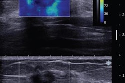
- Explore whether elastography can help guide breast cancer treatment.
- Find out how to build a better breast cancer screening program, according to a presentation delivered at the recent AHRA 2016 meeting in Nashville, TN.
- Is MBI a good alternative for dense breast screening? Definitely, according to research published in the American Journal of Roentgenology.
- Discover what University of Vermont researchers found about the consistency of BI-RADS breast density assessments among radiologists.
- Read what Italian and Australian researchers have to say about the balance between 3D mammography's increased cancer detection and its tendency to boost recall rates.
As always, if you have a comment, report, or article idea to share about any aspect of women's imaging, please contact me.