Dear AuntMinnie Member,
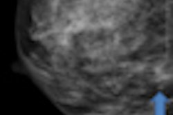
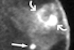
Mammography is burgeoning with new technologies designed to improve its performance in cancer detection. Digital breast tomosynthesis has gotten most of the attention recently, but this week we bring you a story on another intriguing tool: contrast-enhanced dual-energy mammography.
The technique consists of injecting a contrast agent into the patient; then images are acquired at two x-ray energies. The goal is to acquire maps of blood flow in the breast that could detect signs of malignancy in a manner similar to breast MRI -- but at a far lower cost.
Work on the experimental technique was presented at the RSNA meeting earlier this month, and we're featuring a story on the technology in our Women's Imaging Digital Community. Find out how it worked by clicking here.
Coverage of San Antonio symposium
Also in the community, we're featuring coverage of the San Antonio Breast Cancer Symposium, which just wrapped up in Texas.
In one of the presentations, researchers from the Mayo Clinic in Rochester, MN, found that SPECT/CT could reduce the risk of lymphedema in breast cancer patients by enabling radiation oncologists to develop treatment plans that deliver less radiation to critical drainage lymph nodes that are free of disease. Find out how it worked by clicking here.
In another study, Austrian researchers found that among patients undergoing breast conservation surgery in conjunction with radiation therapy, administering a boost of intraoperative electron radiation therapy (IOERT) during surgery and before whole-breast irradiation produced better outcomes than receiving a radiation boost at the end of treatment. That article is available by clicking here.
Get these stories and more in the Women's Imaging Digital Community at women.auntminnie.com.

![A normal mammogram confirmed by three-year radiologic follow-up illustrates reader-marked regions of interest (ROIs) during (A) unaided (round 1) and (B) artificial intelligence (AI)–assisted (round 2) reading. Each colored dot represents an ROI for recall by a human reader. Readers could mark more than one ROI per case, represented by multiple dots of the same color. During AI-assisted reading, the AI system displayed three visible prompts: two with suspicion of malignancy scores of 35% (left mediolateral oblique [L MLO] and craniocaudal [L CC]) and one with a suspicion of malignancy score of 10% (right craniocaudal [R CC]), shown as polygonal overlays. Without AI, six of 10 readers (60%) marked a false-positive ROI. With AI assistance, this fell to two of 10 (20%). R MLO = right mediolateral oblique.](https://img.auntminnie.com/mindful/smg/workspaces/default/uploads/2026/07/2026-07-14-radiology-mammogram-ai-auto-bias.H0bYO8QlWs.jpg?auto=format%2Ccompress&fit=crop&h=100&q=70&w=100)


![A normal mammogram confirmed by three-year radiologic follow-up illustrates reader-marked regions of interest (ROIs) during (A) unaided (round 1) and (B) artificial intelligence (AI)–assisted (round 2) reading. Each colored dot represents an ROI for recall by a human reader. Readers could mark more than one ROI per case, represented by multiple dots of the same color. During AI-assisted reading, the AI system displayed three visible prompts: two with suspicion of malignancy scores of 35% (left mediolateral oblique [L MLO] and craniocaudal [L CC]) and one with a suspicion of malignancy score of 10% (right craniocaudal [R CC]), shown as polygonal overlays. Without AI, six of 10 readers (60%) marked a false-positive ROI. With AI assistance, this fell to two of 10 (20%). R MLO = right mediolateral oblique.](https://img.auntminnie.com/mindful/smg/workspaces/default/uploads/2026/07/2026-07-14-radiology-mammogram-ai-auto-bias.H0bYO8QlWs.jpg?auto=format%2Ccompress&fit=crop&h=112&q=70&w=112)