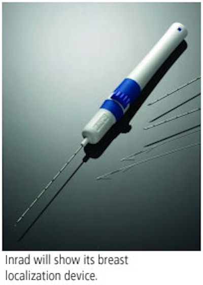
Kentwood, MI-based firm Inrad will feature its UltraWire One-Handed Breast Localization Device at the RSNA show.

The device helps clinicians precisely mark breast lesions using ultrasound guidance. Its design includes a preloaded wire so that the user can place it without having to put down the ultrasound transducer. The needle retracts into the handle after use to reduce needle sticks. The device is available in 5-, 7-, and 9-cm lengths, and will be available in January 2006, according to Inrad.
Also in its booth, Inrad will showcase product line upgrades. The company has added two new lengths to its UltraClip family of tissue markers: the 12-cm length is for clip placement with ultrasound, and the 15-cm length is for clip placements under MRI guidance. Finally, the Ghiatas Beaded Breast Localization Wire for placement with MRI comes with a 20-guage needle with lengths of 5, 7, 9, and 14 cm.
By Kate Madden Yee
AuntMinnie.com contributing writer
November 7, 2005
Copyright © 2005 AuntMinnie.com



![A normal mammogram confirmed by three-year radiologic follow-up illustrates reader-marked regions of interest (ROIs) during (A) unaided (round 1) and (B) artificial intelligence (AI)–assisted (round 2) reading. Each colored dot represents an ROI for recall by a human reader. Readers could mark more than one ROI per case, represented by multiple dots of the same color. During AI-assisted reading, the AI system displayed three visible prompts: two with suspicion of malignancy scores of 35% (left mediolateral oblique [L MLO] and craniocaudal [L CC]) and one with a suspicion of malignancy score of 10% (right craniocaudal [R CC]), shown as polygonal overlays. Without AI, six of 10 readers (60%) marked a false-positive ROI. With AI assistance, this fell to two of 10 (20%). R MLO = right mediolateral oblique.](https://img.auntminnie.com/mindful/smg/workspaces/default/uploads/2026/07/2026-07-14-radiology-mammogram-ai-auto-bias.H0bYO8QlWs.jpg?auto=format%2Ccompress&fit=crop&h=100&q=70&w=100)


![A normal mammogram confirmed by three-year radiologic follow-up illustrates reader-marked regions of interest (ROIs) during (A) unaided (round 1) and (B) artificial intelligence (AI)–assisted (round 2) reading. Each colored dot represents an ROI for recall by a human reader. Readers could mark more than one ROI per case, represented by multiple dots of the same color. During AI-assisted reading, the AI system displayed three visible prompts: two with suspicion of malignancy scores of 35% (left mediolateral oblique [L MLO] and craniocaudal [L CC]) and one with a suspicion of malignancy score of 10% (right craniocaudal [R CC]), shown as polygonal overlays. Without AI, six of 10 readers (60%) marked a false-positive ROI. With AI assistance, this fell to two of 10 (20%). R MLO = right mediolateral oblique.](https://img.auntminnie.com/mindful/smg/workspaces/default/uploads/2026/07/2026-07-14-radiology-mammogram-ai-auto-bias.H0bYO8QlWs.jpg?auto=format%2Ccompress&fit=crop&h=112&q=70&w=112)