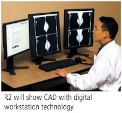
At this year's RSNA show, R2 plans to showcase its ImageChecker DMax CAD system, a high-throughput unit for high-volume mammography centers that accepts film and digital images from all major FFDM products and works with reporting systems from Mammography Reporting System, MagView Information Systems, and PenRad Technologies.

In August, R2 and Linköping, Sweden-based mammography workstation developer Sectra expanded the collaboration they forged in 2004, which integrated R2's CAD technology with the vendor's digital mammography unit. R2 will now sell and support Sectra's Breast Imaging Workstation and Breast PACS, a multimodality, multivendor mammography workstation, to clients in the U.S. and Canada. Sectra's products allow R2's ImageChecker D digital mammography CAD to be used with images from Fischer Imaging, GE Healthcare, Hologic, and Siemens Medical Solutions.
R2 has also updated its CAD algorithm across the ImageChecker product line. Version 8.0 improves false marker rates at any given sensitivity, according to the Sunnyvale, CA-based company.
By Kate Madden Yee
AuntMinnie.com contributing writer
November 7, 2005
Copyright © 2005 AuntMinnie.com



![A normal mammogram confirmed by three-year radiologic follow-up illustrates reader-marked regions of interest (ROIs) during (A) unaided (round 1) and (B) artificial intelligence (AI)–assisted (round 2) reading. Each colored dot represents an ROI for recall by a human reader. Readers could mark more than one ROI per case, represented by multiple dots of the same color. During AI-assisted reading, the AI system displayed three visible prompts: two with suspicion of malignancy scores of 35% (left mediolateral oblique [L MLO] and craniocaudal [L CC]) and one with a suspicion of malignancy score of 10% (right craniocaudal [R CC]), shown as polygonal overlays. Without AI, six of 10 readers (60%) marked a false-positive ROI. With AI assistance, this fell to two of 10 (20%). R MLO = right mediolateral oblique.](https://img.auntminnie.com/mindful/smg/workspaces/default/uploads/2026/07/2026-07-14-radiology-mammogram-ai-auto-bias.H0bYO8QlWs.jpg?auto=format%2Ccompress&fit=crop&h=100&q=70&w=100)


![A normal mammogram confirmed by three-year radiologic follow-up illustrates reader-marked regions of interest (ROIs) during (A) unaided (round 1) and (B) artificial intelligence (AI)–assisted (round 2) reading. Each colored dot represents an ROI for recall by a human reader. Readers could mark more than one ROI per case, represented by multiple dots of the same color. During AI-assisted reading, the AI system displayed three visible prompts: two with suspicion of malignancy scores of 35% (left mediolateral oblique [L MLO] and craniocaudal [L CC]) and one with a suspicion of malignancy score of 10% (right craniocaudal [R CC]), shown as polygonal overlays. Without AI, six of 10 readers (60%) marked a false-positive ROI. With AI assistance, this fell to two of 10 (20%). R MLO = right mediolateral oblique.](https://img.auntminnie.com/mindful/smg/workspaces/default/uploads/2026/07/2026-07-14-radiology-mammogram-ai-auto-bias.H0bYO8QlWs.jpg?auto=format%2Ccompress&fit=crop&h=112&q=70&w=112)