Combining mammography with contrast-enhanced 3-D power Doppler ultrasound may yield an increase in diagnostic accuracy for breast cancer, according to a small study from Thomas Jefferson University in Philadelphia.
"Contrast-enhanced 3-D power Doppler increases the ability to diagnose breast cancer compared to current techniques and may provide a marker of malignancy," said Flemming Forsberg, Ph.D., a professor of radiology and head of research at TJU's Jefferson Ultrasound Institute. He presented the research at the 2002 RSNA meeting in Chicago.
To compare mammography with 2-D and 3-D contrast-enhanced power Doppler for breast cancer diagnosis, a TJU research team studied 55 patients with solid lesions who underwent breast biopsies with histopathological assessment. Of the 55 women, 22 received Levovist (Berlex Laboratories, Wayne, NJ) and 33 received Optison (Mallinckrodt, Hazelwood, MO).
The researchers used an HDI 3000 ultrasound unit (Philips Medical Systems, Andover, MA) to obtain pre-and post-contrast 2-D power Doppler images of the lesion. Three-dimensional data acquisition and reconstruction was performed with an LIS 6000A system (Life Imaging Systems, London, Canada).
Two independent readers blinded to the histopathological results evaluated the images using a six-point scale ranging from "no lesion" to "malignant." The researchers then performed logistic regression analysis to combine mammographic and ultrasound findings, and computed the area under the ROC curve for each modality individually as well as in combination for mammography and 2-D and 3-D ultrasound (both pre-and-post contrast), Forsberg said. The study team also compared histopathological variables and imaging parameters using Mann-Whitney statistics.
Mammography findings were available for 50 subjects, while biopsy was available in 54 cases, 2-D ultrasound in 53, and 3-D ultrasound in 52 patients. Of the 50 subjects with all four measures, 15 (30%) had malignant breast lesions.
 |
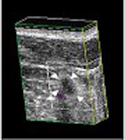
Ductal carcinoma in situ (arrows) shown in 3-D power Doppler before (above) and after (below) injection of 1 ml of Optison. Images courtesy of Flemming Forsberg, Ph.D.
 |
The area under the ROC curve for breast cancer diagnosis was 0.51 for 2-D contrast, 0.6 for 3-D ultrasound, and 0.76 for 3-D contrast. Mammography yielded an area under the ROC curve of 0.86, which grew to 0.88 when combined with 2-D contrast, 0.9 when combined with 3-D contrast, and 0.96 when combined with all ultrasound modalities, Forsberg said.
"When you combine mammography with ultrasound, it's usually better than mammography alone, based on the experience (of this patient population)," he said.
The histopathological diagnosis of benign or malignant had a statistically significant correlation with the presence or absence of anastamoses assessed with contrast-enhanced 3-D power Doppler, and with the degree of vascularity seen with 3-D contrast ultrasound, Forsberg said.
"Only the 3-D power Doppler seems to give a marker of the malignancy," Forsberg said. "Moreover, the 3-D signals were more likely to be deemed acceptable."
By Erik L. RidleyAuntMinnie.com staff writer
April 11, 2003
Related Reading
New adjunctive breast techniques improve positive biopsy rates, February 25, 2003
Mammo plus US negates need for breast MR, December 23, 2002
Breast Ultrasound: A Systemic Approach to Technique and Image Interpretation, December 23, 2002
The Practice of Breast Ultrasound, December 23, 2002
ARRT plans certification in breast ultrasound, December 20, 2002
Copyright © 2003 AuntMinnie.com