Contrast-enhanced mammography (CEM) may be superior to low-energy image alone in diagnosing ductal carcinoma in situ (DCIS), suggest findings published May 22 in Clinical Radiology.
CEM showed higher sensitivity over low-energy imaging regardless of pathological nuclear grading or the presence of calcifications, wrote a team led by Liping Wang, MD, from the Affiliated Hospital of Qingdao University in Yantai, Shandong, China.
“CEM could be a good choice for the preoperative diagnosis and prognostic evaluation of DCIS,” Wang and colleagues wrote.
Some DCIS cases may progress to invasive cancer, making early detection and reliable prognosis prediction important for treatment strategy. Previous studies suggest that pathological nuclear grading is a key prognostic factor of DCIS.
The researchers called CEM’s application in breast disease diagnosis a “current hotspot,” although they also noted a lack of data on CEM and pathological nuclear grades of DCIS.
Wang and colleagues studied CEM’s performance in diagnosing DCIS as well as correlations between DCIS manifestations on CEM and pathological nuclear grades of DCIS.
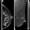
The single-center retrospective study included 190 women with pure DCIS who underwent CEM within two weeks preoperatively. Final analysis included CEM images of 196 DCIS lesions (bilateral DCIS in six women).
CEM showed significantly higher sensitivity compared to low-energy imaging alone, regardless of the presence of calcifications and pathological nuclear grading.
Sensitivity comparison between CEM, low-energy imaging | |||
DCIS case | Low-energy imaging | CEM | P-value |
Overall | 72.4% | 89.3% | < 0.001 |
Calcified | 91.2% | 96.8% | 0.016 |
Non-calcified | 39.4% | 76.1% | < 0.001 |
Low-grade | 61.1% | 83.3% | 0.008 |
Intermediate-grade | 65.8% | 87.3% | < 0.001 |
High-grade | 84.0% | 93.8% | 0.008 |
CEM also proved to be equally sensitive in diagnosing different grades of DCIS (p = 0.183), with CEM being superior to low-energy image alone in showing the extent of DCIS lesions, the researchers wrote.
The team also highlighted high-grade DCIS showing more calcifications and larger size than low-grade DCIS on CEM and showing higher enhancement conspicuity than low- and intermediate-grade DCIS (p < 0.05). And low-grade DCIS showed less pleomorphic and fine linear branching calcifications than intermediate- and high-grade DCIS (p < 0.05).
Finally, the team noted no significant differences between the three grading groups in terms of enhancement percentage and enhancement morphology (p > 0.05).
The study authors highlighted that clinicians could better tailor treatment strategies to individual women with CEM improving the detection rate of all nuclear grades of DCIS.
Read the full study here.


![A normal mammogram confirmed by three-year radiologic follow-up illustrates reader-marked regions of interest (ROIs) during (A) unaided (round 1) and (B) artificial intelligence (AI)–assisted (round 2) reading. Each colored dot represents an ROI for recall by a human reader. Readers could mark more than one ROI per case, represented by multiple dots of the same color. During AI-assisted reading, the AI system displayed three visible prompts: two with suspicion of malignancy scores of 35% (left mediolateral oblique [L MLO] and craniocaudal [L CC]) and one with a suspicion of malignancy score of 10% (right craniocaudal [R CC]), shown as polygonal overlays. Without AI, six of 10 readers (60%) marked a false-positive ROI. With AI assistance, this fell to two of 10 (20%). R MLO = right mediolateral oblique.](https://img.auntminnie.com/mindful/smg/workspaces/default/uploads/2026/07/2026-07-14-radiology-mammogram-ai-auto-bias.H0bYO8QlWs.jpg?auto=format%2Ccompress&dpr=2&fit=crop&h=167&q=70&w=250)