Resting-state functional MRI (fMRI) scans show measurable differences in brain connectivity across menopause stages, with postmenopausal women displaying significantly reduced neural communication compared to premenopausal women, researchers have reported.
The findings suggest that menopause represents "an important neurological transition that may influence both cognitive experiences in the present and long-term brain aging," according to Julie Dumas, PhD, and colleague Abigail Testo, PhD, both of the University of Vermont in Burlington. The study results were published June 9 in Menopause.
"With decades of life remaining after menopause, it is important to understand the neurological effects of hormone changes at midlife," Testo said in a statement released by the university. "Our research contributes to the growing body of work examining the relationship between menopause and the brain."
According to the National Institutes of Health, approximately 6,000 women enter menopause each day in the United States, or 1.3 million annually, Dumas and Testo noted. They sought to assess the relationship between menopause stage and resting-state functional connectivity during midlife via a study that included data taken from the Human Connectome Project - Aging 2.0 regarding 151 women between the ages of 40 and 55.
All study participants underwent an fMRI exam; Dumas and Testo used Conn Toolbox to evaluate the strength of functional associations between brain regions at rest at both specific brain region and brain region cluster levels (Conn Toolbox is software used to compute, display, and analyze functional connectivity from brain fMRI sequences.)
Dumas and Testo reported the following:
- Differences in resting-state functional connectivity between the supramarginal gyrus, right anterior division, and right planum temporale at the connection level were identified between participants in the pre-, peri-, and postmenopausal groups.
- Comparison of the pre- and postmenopausal groups showed one cluster of altered resting-state connectivity that was lower in the postmenopausal group compared to the premenopausal group; these regions included the left and right supramarginal gyrus, the anterior division, and the right and left planum temporale.
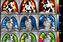
 Network-level analysis comparing functional association strength between ROIs of pre- and postmenopausal groups. Lower levels of connectivity were identified among one cluster (F3,106 = 8.84, Puncorrected < 0.001, PFDR = 0.012) in the postmenopausal group, which included the right supramarginal gyrus anterior division, right planum temporale, and right Heschl’s gyrus, among others. FDR, false discovery rate; ROI, region of interest.Menopause
These differences appear to be linked to hormonal changes, particularly fluctuations in estrogen, one of the most essential hormones that play an integral role in sexual and reproductive development in women, they explained.
"Further research is needed to determine the role that differences in brain functioning before and after menopause play in individual differences in the experience of aging," Dumas and Testo concluded.
Access the full study here.