Dear AuntMinnie Member,
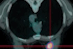
Performing PET/CT for breast imaging at two points in time improves the technique's accuracy compared to studies done at just one time point. But even with this improvement, dual-time-point PET/CT may not be as good as breast MRI for small lesions.
MRI remains the preferred modality for characterizing those lesions, according to an article by staff writer Kate Madden Yee that we're featuring this week in our Women's Imaging Digital Community. Italian researchers compared FDG-PET/CT taken three hours apart to contrast-enhanced breast MRI, which has been winning favor as a method for analyzing suspicious lesions found on mammography.
The researchers found that the time difference between PET/CT studies improved the technique's sensitivity and specificity by over 10 percentage points. But the modality still fell short of the accuracy produced by breast MRI. Get the rest of the story by clicking here.
In other news, staff writer Cynthia Keen reports on another group of Italian researchers who found that electron-beam intraoperative radiation therapy (IORT) can be performed on pregnant women undergoing breast-conservation surgery, without the risk of delivering too much radiation to the fetus. That story is available by clicking here.
Get these stories and more in our Women's Imaging Digital Community, at women.auntminnie.com.