CHICAGO - Combining parameters from 2D shear-wave elastography can help assess hepatic characteristics in patients with biopsy-proven nonalcoholic fatty liver disease, suggests research shown at the RSNA annual meeting.
Dr. Katsutoshi Sugimoto, PhD, from Tokyo Medical Hospital in Japan presented the study's results, which demonstrated high performance in helping accurately diagnose nonalcoholic steatohepatitis.
"Dispersion slope, attenuation coefficient, and shear-wave speed were ultrasound parameters that may help estimate inflammation, steatosis, and fibrosis. They may allow for noninvasive diagnosis of nonalcoholic steatohepatitis in a single examination," Sugimoto said.
Previous research led by Sugimoto et al demonstrated that combining the aforementioned parameters can accomplish this goal. However, they noted that prospective multicenter study designs and larger, more diverse cohorts are needed to validate these findings. Other research has previously demonstrated the promise of ultrasound in liver imaging as an alternative to biopsy.
Sugimoto and colleagues since their initial findings created a global multicenter study called the Innovative Liver Elasticity, Attenuation, and Dispersion Ultrasound Study (iLead). In his presentation, Sugimoto showed the interim results of the study.
The team wanted to find out the diagnostic performance of dispersion slope, attenuation coefficient, and shear-wave speed measures to assess patients just before they underwent liver biopsy.
The researchers looked at data from 74 patients with an average age of 53 years. Out of these, 35 were male and 39 were female. Also, 48 were Asian, 22 were white, and four were LatinX.
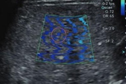
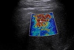
 Researchers in Tokyo presented results of a study examining the potential of ultrasonic parameters on 2D shear-wave elastography in diagnosing biopsy-proven nonalcoholic fatty liver disease. The results were shown at the RSNA annual meeting. Here, shear-wave speed and dispersion slope are compared in assessing lobular inflammation. Image courtesy of Dr. Katsutoshi Sugimoto.
The researchers found that the combination of all three parameters showed an area under the curve (AUC) of 0.791 for the diagnosis of nonalcoholic steatohepatitis.
Individually, the parameters showed variable performance. For example, dispersion slope had an AUC of 0.696 for identifying moderate lobular inflammation and 0.920 for identifying marked inflammation. Shear-wave speed meanwhile yielded superior performance for moderate (AUC, 0.708) and lower-grade inflammation (AUC, 0.779). For marked inflammation, shear-wave speed had an AUC of 0.917.
The attenuation coefficient had high performance for identifying steatosis. This included an AUC of 0.857 for moderate steatosis and 0.857 for severe steatosis.
Finally, shear-wave speed allowed researchers to identify fibrosis with high performance. This included AUCs of 0.892 for stage II fibrosis, 0.886 for stage III, and 0.845 for stage IV.
Sugimoto said the team should collect more information regarding liver biopsy to further validate their results.