A new telediagnostic ultrasound protocol that doesn't need a trained sonographer or high-speed internet showed success in a pilot program held in Peru. The study was presented at this month's virtual American Roentgen Ray Society (ARRS) meeting.
Researchers from the University of Rochester developed a scanning protocol that could be performed by staff in the field with minimal training, with images sent over low-bandwidth connections for interpretation by experts. Imaging readers reported a high level of satisfaction with the images.
"For the majority of the world that lacks access to diagnostic imaging, this could really improve the health of the global community by giving them access," said Dr. Thomas Marini from the University of Rochester.
About two-thirds of the world doesn't have access to diagnostic imaging. While ultrasound could be deployed into rural areas, hindrances such as lack of trained sonographers, high-speed internet for teleconferencing, and healthcare infrastructure exist.
In his ARRS talk, Marini described a system designed to overcome these challenges. Here's how it works: Operators without prior ultrasound experience use a protocol the authors called volume sweep imaging to scan across preset areas of the patient's body in straight lines, with areas of interest based on external body landmarks. The operators do not interpret imaging videos or adjust an ultrasound machine.
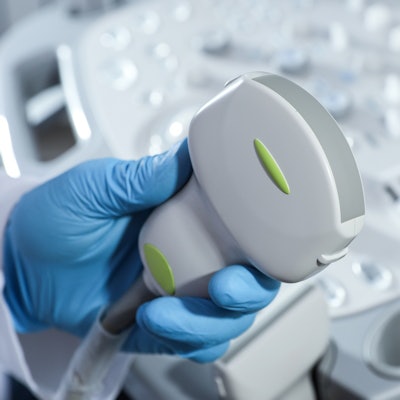
The sweeps are captured by a portable ultrasound scanner that's connected to a tablet computer, which saves the data as video files and also records patient information. The files are then compressed to a size that can successfully travel over a low bandwidth to a cloud storage system in less than five minutes.
From there, a radiologist downloads the images and information from the cloud and can interpret them the same way they would for any other imaging study. The results are sent back to the tablet and then to the patient.
"There's minimal technical skill involved [for operators]," Marini said. "Anybody can really learn how to do this, as long as they can perform basic motions of the probe."
In his ARRS talk, Marini described how operators without prior ultrasound experience were trained for a total of nine hours on volume sweep imaging protocols. They were then sent to scan 67 study participants in Peru, and images were sent to the U.S. for interpretation.
Out of the participants, 29 underwent the right upper quadrant protocol, 22 underwent the thyroid protocol, and 16 underwent the obstetrics protocol. Blinded expert readers interpreted the volume sweep imaging acquisitions using standardized sheets detailing visualization of major structures, their confidence in estimations, and image quality.
The readers determined that features such as thyroid cysts, fetuses, and amniotic fluid were all clearly identified in the participants. In all, 96% of the scans were judged to have "acceptable" or "excellent" imaging quality.
"Many of these [images] look like they could be obtained by a sonographer," Marini said.
He said a public health study is needed to identify where this system fits into the healthcare environment. Future use of the system is possible for other protocols such as the breast and lung, he added.



![Examples of ultrasound findings and techniques. (A) Images in a 39-year-old male patient with a mass in the left thigh. The mass is heterogeneous on the B-mode US image (compared with the patient in D) and showed increased microvascularity (superb microvascular imaging [SMI]) and shear-wave elastography (SWE) values. Undifferentiated pleomorphic sarcoma was diagnosed at biopsy (with pleomorphic rhabdomyosarcoma in surgical specimen). (B) Images in an 18-year-old male patient with a mass in the left leg. The mass is hypoechoic on the B-mode image, with no other findings suggestive of malignancy. The lesion is in contact with the cortex of the tibia, which is slightly irregular. CT revealed a doubtful anteromedial tibial erosion. The microvascular study demonstrated high vascularization, suggestive of malignancy. Periosteal Ewing sarcoma was diagnosed with both histologic and immunohistochemical confirmation. (C) Images in a 69-year-old female patient with a lump growing on the outside of the left leg. Multiple SWE examinations were performed (please note the high values obtained in the measurements, whereas the color map highlights the stiffness relative to adjacent tissues). SMI showed areas of increased vascularization to target for sampling. Undifferentiated spindle cell sarcoma was diagnosed at biopsy, with residual leiomyosarcoma in the surgical specimen after neoadjuvant therapy. (D) Images in a 56-year-old female patient with a mass in the right thigh. The mass is heterogeneous at both B-mode ultrasound (similar to patient A) and MRI (coronal T2-weighted spectral attenuated inversion recovery [SPAIR]; T1-weighted pre-contrast and postcontrast imaging), which even shows uptake after the administration of paramagnetic contrast material, which is traditionally suggestive of malignancy. Low values at SMI and elastography are suggestive of benignity. Spindle cell lipoma was diagnosed at biopsy, with atypical spindle cell lipomatous tumor in the surgical specimen.](https://img.auntminnie.com/mindful/smg/workspaces/default/uploads/2026/08/images-radiol250278fig2.APCFLSvX6p.jpg?auto=format%2Ccompress&fit=crop&h=100&q=70&w=100)


![Examples of ultrasound findings and techniques. (A) Images in a 39-year-old male patient with a mass in the left thigh. The mass is heterogeneous on the B-mode US image (compared with the patient in D) and showed increased microvascularity (superb microvascular imaging [SMI]) and shear-wave elastography (SWE) values. Undifferentiated pleomorphic sarcoma was diagnosed at biopsy (with pleomorphic rhabdomyosarcoma in surgical specimen). (B) Images in an 18-year-old male patient with a mass in the left leg. The mass is hypoechoic on the B-mode image, with no other findings suggestive of malignancy. The lesion is in contact with the cortex of the tibia, which is slightly irregular. CT revealed a doubtful anteromedial tibial erosion. The microvascular study demonstrated high vascularization, suggestive of malignancy. Periosteal Ewing sarcoma was diagnosed with both histologic and immunohistochemical confirmation. (C) Images in a 69-year-old female patient with a lump growing on the outside of the left leg. Multiple SWE examinations were performed (please note the high values obtained in the measurements, whereas the color map highlights the stiffness relative to adjacent tissues). SMI showed areas of increased vascularization to target for sampling. Undifferentiated spindle cell sarcoma was diagnosed at biopsy, with residual leiomyosarcoma in the surgical specimen after neoadjuvant therapy. (D) Images in a 56-year-old female patient with a mass in the right thigh. The mass is heterogeneous at both B-mode ultrasound (similar to patient A) and MRI (coronal T2-weighted spectral attenuated inversion recovery [SPAIR]; T1-weighted pre-contrast and postcontrast imaging), which even shows uptake after the administration of paramagnetic contrast material, which is traditionally suggestive of malignancy. Low values at SMI and elastography are suggestive of benignity. Spindle cell lipoma was diagnosed at biopsy, with atypical spindle cell lipomatous tumor in the surgical specimen.](https://img.auntminnie.com/mindful/smg/workspaces/default/uploads/2026/08/images-radiol250278fig2.APCFLSvX6p.jpg?auto=format%2Ccompress&fit=crop&h=112&q=70&w=112)