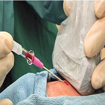
Placing arterial lines under the guidance of point-of-care ultrasound (POCUS) outperformed traditional landmark guidance in research presented Tuesday at the virtual 2021 American Institute of Ultrasound in Medicine (AIUM) annual meeting.
Research presented by Dr. Ryan Gibbons from Temple University showed that ultrasound guidance led to fewer complications, fewer attempts, and less placement time for interns inserting arterial lines in patients in emergency departments. Gibbons presented findings originally published in September 2020 in the Journal of Emergency Medicine.
Prior to point-of-care ultrasound, landmark guidance was considered the standard of care, but for up to 30% of patients, anatomic landmarks are unreliable, leading to a significant number of complications, Gibbons said. While major complications from landmark guidance are rare, they can lead to vascular injury.
"Furthermore, patient body habitus and hemodynamic instability significantly limit the accuracy of palpation," Gibbons told AuntMinnie.com.
At AIUM 2021, Gibbons discussed results from a randomized controlled trial of 40 patients at Temple University Hospital: 20 patients received ultrasound guidance with a POCUS device, while the other 20 received landmark guidance. The groups were similar in age, sex, and body mass index.
Eighteen novice emergency medicine interns with fewer than 15 previous placements performed the cannulation. They were limited to three attempts or 15 minutes without access and then were instructed to cross over to the other technique.
All 20 patients who received ultrasound guidance had successful cannulation. Meanwhile, only three patients who received landmark guidance saw similar success.
| POCUS guidance vs. landmark guidance for arterial placement | |||
| POCUS guidance | Landmark guidance | p-value | |
| First-pass success rate | 75% | 0% | < 0.0001 |
| Overall success | 100% | 15% | < 0.0001 |
| Attempts (mean) | 1.30 | 2.95 | < 0.00001 |
| Time of placement (mean) | 264 seconds | 524 seconds | 0.00424 |
Some patients with multiple failed attempts at landmark guidance were crossed over to ultrasound guidance; POCUS was 100% successful in these individuals, with a mean of 1.29 attempts and 170 seconds for placement.
Ultrasound guidance proved to be superior to traditional landmark guidance in a randomized trial conducted by researchers at Temple University. Image courtesy of Dr. Ryan Gibbons.Limitations for the study included patients being unblinded to the fact that the trial was conducted by the Division of Ultrasound, as well as the trial taking place in one center. The researchers also did not account for significant prior point-of-care ultrasound experience.
Gibbons told AuntMinnie.com the team is completing a study comparing ultrasound guidance with landmark guidance in arthrocentesis of medium joints, as well as multicenter studies looking at the role of ultrasound in COVID-19 and cardiac arrest.


![Examples of ultrasound findings and techniques. (A) Images in a 39-year-old male patient with a mass in the left thigh. The mass is heterogeneous on the B-mode US image (compared with the patient in D) and showed increased microvascularity (superb microvascular imaging [SMI]) and shear-wave elastography (SWE) values. Undifferentiated pleomorphic sarcoma was diagnosed at biopsy (with pleomorphic rhabdomyosarcoma in surgical specimen). (B) Images in an 18-year-old male patient with a mass in the left leg. The mass is hypoechoic on the B-mode image, with no other findings suggestive of malignancy. The lesion is in contact with the cortex of the tibia, which is slightly irregular. CT revealed a doubtful anteromedial tibial erosion. The microvascular study demonstrated high vascularization, suggestive of malignancy. Periosteal Ewing sarcoma was diagnosed with both histologic and immunohistochemical confirmation. (C) Images in a 69-year-old female patient with a lump growing on the outside of the left leg. Multiple SWE examinations were performed (please note the high values obtained in the measurements, whereas the color map highlights the stiffness relative to adjacent tissues). SMI showed areas of increased vascularization to target for sampling. Undifferentiated spindle cell sarcoma was diagnosed at biopsy, with residual leiomyosarcoma in the surgical specimen after neoadjuvant therapy. (D) Images in a 56-year-old female patient with a mass in the right thigh. The mass is heterogeneous at both B-mode ultrasound (similar to patient A) and MRI (coronal T2-weighted spectral attenuated inversion recovery [SPAIR]; T1-weighted pre-contrast and postcontrast imaging), which even shows uptake after the administration of paramagnetic contrast material, which is traditionally suggestive of malignancy. Low values at SMI and elastography are suggestive of benignity. Spindle cell lipoma was diagnosed at biopsy, with atypical spindle cell lipomatous tumor in the surgical specimen.](https://img.auntminnie.com/mindful/smg/workspaces/default/uploads/2026/08/images-radiol250278fig2.APCFLSvX6p.jpg?auto=format%2Ccompress&fit=crop&h=100&q=70&w=100)


![Examples of ultrasound findings and techniques. (A) Images in a 39-year-old male patient with a mass in the left thigh. The mass is heterogeneous on the B-mode US image (compared with the patient in D) and showed increased microvascularity (superb microvascular imaging [SMI]) and shear-wave elastography (SWE) values. Undifferentiated pleomorphic sarcoma was diagnosed at biopsy (with pleomorphic rhabdomyosarcoma in surgical specimen). (B) Images in an 18-year-old male patient with a mass in the left leg. The mass is hypoechoic on the B-mode image, with no other findings suggestive of malignancy. The lesion is in contact with the cortex of the tibia, which is slightly irregular. CT revealed a doubtful anteromedial tibial erosion. The microvascular study demonstrated high vascularization, suggestive of malignancy. Periosteal Ewing sarcoma was diagnosed with both histologic and immunohistochemical confirmation. (C) Images in a 69-year-old female patient with a lump growing on the outside of the left leg. Multiple SWE examinations were performed (please note the high values obtained in the measurements, whereas the color map highlights the stiffness relative to adjacent tissues). SMI showed areas of increased vascularization to target for sampling. Undifferentiated spindle cell sarcoma was diagnosed at biopsy, with residual leiomyosarcoma in the surgical specimen after neoadjuvant therapy. (D) Images in a 56-year-old female patient with a mass in the right thigh. The mass is heterogeneous at both B-mode ultrasound (similar to patient A) and MRI (coronal T2-weighted spectral attenuated inversion recovery [SPAIR]; T1-weighted pre-contrast and postcontrast imaging), which even shows uptake after the administration of paramagnetic contrast material, which is traditionally suggestive of malignancy. Low values at SMI and elastography are suggestive of benignity. Spindle cell lipoma was diagnosed at biopsy, with atypical spindle cell lipomatous tumor in the surgical specimen.](https://img.auntminnie.com/mindful/smg/workspaces/default/uploads/2026/08/images-radiol250278fig2.APCFLSvX6p.jpg?auto=format%2Ccompress&fit=crop&h=112&q=70&w=112)