A scoring system based on lung ultrasound scans could predict intubation, death, and other negative outcomes in COVID-19 patients, according to a recent study published in Annals of Intensive Care.
The study evaluated a disease score threshold using portable ultrasound for assessing COVID-19 patients admitted to the emergency department at Hospital das Clínicas da Faculdade de Medicina da Universidade de São Paulo in Brazil. Results were reported by Dr. Julio Cesar de Alencar and colleagues.
Lung ultrasound scores are semiquantitative scoring systems that measure lung aeration loss caused by different pathologies. Previous studies have found the scores to be useful in assessing the severity of disease in patients with COVID-19, while other research has found that the scores can predict the need for mechanical ventilation and patient mortality.
In the current study, researchers concluded that lung ultrasound scores were associated with a likelihood of death -- the primary outcome in the study -- as well as a range of other important outcomes: the extent of parenchymal involvement as seen on chest CT, endotracheal intubation, and admission to intensive care units (ICUs). All of these results were statistically significant.
The authors noted that lung ultrasound is an attractive and useful alternative to chest CT, which is sensitive for assessment of disease severity and for guiding clinical management, but which has a number of downsides like exposure to radiation, the need for rigorous infection control, and lack of availability in developing countries.
"Recent reports suggest that, in COVID-19 patients, lung ultrasound could be useful in several scenarios: to quantify the severity of lung involvement in periodic assessments, to look for findings suggestive of pneumonia, and to monitor the dynamic effects of mechanical ventilation and recruitment maneuvers on lung aeration," the authors wrote.
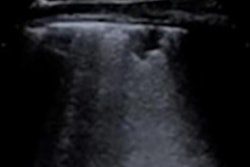
For the study, researchers used a portable ultrasound system (SonoSite Edge II, Fujifilm SonoSite) to examine 12 regions of the lung in 180 patients admitted to the emergency room and determined a semiquantitative score that measures lung aeration. The imaging study took only five minutes to complete, the authors noted.
The researchers then calculated the ability of the score to predict various patient outcomes, based on a cutoff value.
| Ability of lung ultrasound score to predict patient outcomes | |||
| Lung ultrasound score cutoff | Sensitivity | Specificity | |
| Discharged alive | < 16 | 40% | 90% |
| Deceased | > 26 | 23% | 90% |
| Not intubated during hospital stay | < 15 | 41% | 90% |
| Intubated during hospital stay | ≥ 25 | 27% | 93% |
| Not admitted to ICU | < 13 | 31% | 91% |
| Admitted to ICU | ≥ 25 | 22% | 93% |
"The COVID-19 pandemic has brought numerous patients complaining of fever, cough, and dyspnea to [emergency departments] around the world," the authors wrote. "Proper assessment of the severity and extent of pulmonary involvement is of paramount importance to select patients who will be admitted to hospital wards or ICUs and thus ensure adequate management of overwhelmed healthcare resources."

![Examples of ultrasound findings and techniques. (A) Images in a 39-year-old male patient with a mass in the left thigh. The mass is heterogeneous on the B-mode US image (compared with the patient in D) and showed increased microvascularity (superb microvascular imaging [SMI]) and shear-wave elastography (SWE) values. Undifferentiated pleomorphic sarcoma was diagnosed at biopsy (with pleomorphic rhabdomyosarcoma in surgical specimen). (B) Images in an 18-year-old male patient with a mass in the left leg. The mass is hypoechoic on the B-mode image, with no other findings suggestive of malignancy. The lesion is in contact with the cortex of the tibia, which is slightly irregular. CT revealed a doubtful anteromedial tibial erosion. The microvascular study demonstrated high vascularization, suggestive of malignancy. Periosteal Ewing sarcoma was diagnosed with both histologic and immunohistochemical confirmation. (C) Images in a 69-year-old female patient with a lump growing on the outside of the left leg. Multiple SWE examinations were performed (please note the high values obtained in the measurements, whereas the color map highlights the stiffness relative to adjacent tissues). SMI showed areas of increased vascularization to target for sampling. Undifferentiated spindle cell sarcoma was diagnosed at biopsy, with residual leiomyosarcoma in the surgical specimen after neoadjuvant therapy. (D) Images in a 56-year-old female patient with a mass in the right thigh. The mass is heterogeneous at both B-mode ultrasound (similar to patient A) and MRI (coronal T2-weighted spectral attenuated inversion recovery [SPAIR]; T1-weighted pre-contrast and postcontrast imaging), which even shows uptake after the administration of paramagnetic contrast material, which is traditionally suggestive of malignancy. Low values at SMI and elastography are suggestive of benignity. Spindle cell lipoma was diagnosed at biopsy, with atypical spindle cell lipomatous tumor in the surgical specimen.](https://img.auntminnie.com/mindful/smg/workspaces/default/uploads/2026/08/images-radiol250278fig2.APCFLSvX6p.jpg?auto=format%2Ccompress&fit=crop&h=100&q=70&w=100)


![Examples of ultrasound findings and techniques. (A) Images in a 39-year-old male patient with a mass in the left thigh. The mass is heterogeneous on the B-mode US image (compared with the patient in D) and showed increased microvascularity (superb microvascular imaging [SMI]) and shear-wave elastography (SWE) values. Undifferentiated pleomorphic sarcoma was diagnosed at biopsy (with pleomorphic rhabdomyosarcoma in surgical specimen). (B) Images in an 18-year-old male patient with a mass in the left leg. The mass is hypoechoic on the B-mode image, with no other findings suggestive of malignancy. The lesion is in contact with the cortex of the tibia, which is slightly irregular. CT revealed a doubtful anteromedial tibial erosion. The microvascular study demonstrated high vascularization, suggestive of malignancy. Periosteal Ewing sarcoma was diagnosed with both histologic and immunohistochemical confirmation. (C) Images in a 69-year-old female patient with a lump growing on the outside of the left leg. Multiple SWE examinations were performed (please note the high values obtained in the measurements, whereas the color map highlights the stiffness relative to adjacent tissues). SMI showed areas of increased vascularization to target for sampling. Undifferentiated spindle cell sarcoma was diagnosed at biopsy, with residual leiomyosarcoma in the surgical specimen after neoadjuvant therapy. (D) Images in a 56-year-old female patient with a mass in the right thigh. The mass is heterogeneous at both B-mode ultrasound (similar to patient A) and MRI (coronal T2-weighted spectral attenuated inversion recovery [SPAIR]; T1-weighted pre-contrast and postcontrast imaging), which even shows uptake after the administration of paramagnetic contrast material, which is traditionally suggestive of malignancy. Low values at SMI and elastography are suggestive of benignity. Spindle cell lipoma was diagnosed at biopsy, with atypical spindle cell lipomatous tumor in the surgical specimen.](https://img.auntminnie.com/mindful/smg/workspaces/default/uploads/2026/08/images-radiol250278fig2.APCFLSvX6p.jpg?auto=format%2Ccompress&fit=crop&h=112&q=70&w=112)