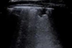
The findings on initial lung scans with point-of-care ultrasound (POCUS) can predict which patients with COVID-19 are at a greater risk of death, according to a prospective study from Italy published on July 15 in Ultrasound in Medicine & Biology.
Physicians in Rome performed lung ultrasound scans on 41 adult patients who visited a tertiary emergency department with symptoms of the novel coronavirus disease. Patients who later died or were later admitted to the intensive care unit (ICU) had significantly worse pathological findings on their initial scan.
"Our study shows that [lung ultrasound scan] is able to detect COVID-19 pneumonia and to predict, during the first evaluation in the emergency department, patients at risk of intensive care unit admission and death," wrote the authors, led by Nicola Bonadia, from the department of emergency medicine at Agostino Gemelli University Policlinic.
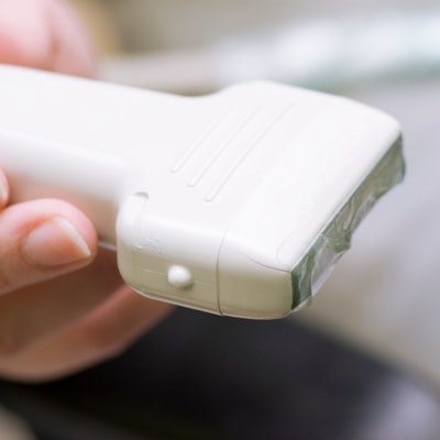
Physicians performed point-of-care ultrasound (POCUS) scans on all patients with suspected cases of COVID-19 who visited the emergency department in March. The emergency department staff used a pocket device with a wireless 6-MHz convex probe and followed a previously described lung ultrasound protocol that includes 14 chest areas.
Each scanned area received a numeric score of 0 to 3 based on the severity of the findings in that section. A higher lung ultrasound score (LUS) signified worse disease severity, with a score of 3 indicating dense or large areas of white lung with or without subpleural consolidations.
The authors analyzed the lung ultrasound findings from 41 patients with a positive SARS-CoV-2 test result and known outcomes. They specifically excluded children and patients with less than six months life expectancy due to preexisting chronic conditions, such as advanced cancer or dementia.
More than 90% of adult patients with COVID-19 had at least one area with an abnormal lung ultrasound finding. Pathological findings occurred in all 14 scanned areas but were most prominent in the lateral lung regions, the authors noted.

Patients with fatal cases of COVID-19 had pathological findings in 100% of scanned areas, compared with just 50% of scanned areas in discharged patients. These patients also had a mean lung ultrasound score of 1.43, compared with 1.0 in discharged cases.
Similarly, patients admitted to the ICU had pathological findings in 93% of scanned areas, compared with just 20% of areas in patients not admitted to the ICU. ICU patients also had a mean LUS of 1.36, compared with 1.0 in non-ICU patients.
Based on their findings, the authors determined the cutoff for a strict definition of COVID-19 pneumonia should be an LUS of 0.4 and a pathological findings rate of at least 20%. Furthermore, no study participants died if they had a mean LUS less than 1.1 and an average pathological rate under 70%.
The authors cautioned their study took place at one institution and had a small sample size. Nevertheless, the findings may help health professionals better triage patients with COVID-19 and spur future studies to evaluate whether lung ultrasound scores can guide patient treatment and admission decisions.
"We found a significant correlation between ultrasound findings and severity of the disease, assessed as mortality and need for ICU admission," the authors wrote. "To our knowledge, this is the first study describing the predictive role of LUS in patients with COVID-19."


![Examples of ultrasound findings and techniques. (A) Images in a 39-year-old male patient with a mass in the left thigh. The mass is heterogeneous on the B-mode US image (compared with the patient in D) and showed increased microvascularity (superb microvascular imaging [SMI]) and shear-wave elastography (SWE) values. Undifferentiated pleomorphic sarcoma was diagnosed at biopsy (with pleomorphic rhabdomyosarcoma in surgical specimen). (B) Images in an 18-year-old male patient with a mass in the left leg. The mass is hypoechoic on the B-mode image, with no other findings suggestive of malignancy. The lesion is in contact with the cortex of the tibia, which is slightly irregular. CT revealed a doubtful anteromedial tibial erosion. The microvascular study demonstrated high vascularization, suggestive of malignancy. Periosteal Ewing sarcoma was diagnosed with both histologic and immunohistochemical confirmation. (C) Images in a 69-year-old female patient with a lump growing on the outside of the left leg. Multiple SWE examinations were performed (please note the high values obtained in the measurements, whereas the color map highlights the stiffness relative to adjacent tissues). SMI showed areas of increased vascularization to target for sampling. Undifferentiated spindle cell sarcoma was diagnosed at biopsy, with residual leiomyosarcoma in the surgical specimen after neoadjuvant therapy. (D) Images in a 56-year-old female patient with a mass in the right thigh. The mass is heterogeneous at both B-mode ultrasound (similar to patient A) and MRI (coronal T2-weighted spectral attenuated inversion recovery [SPAIR]; T1-weighted pre-contrast and postcontrast imaging), which even shows uptake after the administration of paramagnetic contrast material, which is traditionally suggestive of malignancy. Low values at SMI and elastography are suggestive of benignity. Spindle cell lipoma was diagnosed at biopsy, with atypical spindle cell lipomatous tumor in the surgical specimen.](https://img.auntminnie.com/mindful/smg/workspaces/default/uploads/2026/08/images-radiol250278fig2.APCFLSvX6p.jpg?auto=format%2Ccompress&fit=crop&h=100&q=70&w=100)


![Examples of ultrasound findings and techniques. (A) Images in a 39-year-old male patient with a mass in the left thigh. The mass is heterogeneous on the B-mode US image (compared with the patient in D) and showed increased microvascularity (superb microvascular imaging [SMI]) and shear-wave elastography (SWE) values. Undifferentiated pleomorphic sarcoma was diagnosed at biopsy (with pleomorphic rhabdomyosarcoma in surgical specimen). (B) Images in an 18-year-old male patient with a mass in the left leg. The mass is hypoechoic on the B-mode image, with no other findings suggestive of malignancy. The lesion is in contact with the cortex of the tibia, which is slightly irregular. CT revealed a doubtful anteromedial tibial erosion. The microvascular study demonstrated high vascularization, suggestive of malignancy. Periosteal Ewing sarcoma was diagnosed with both histologic and immunohistochemical confirmation. (C) Images in a 69-year-old female patient with a lump growing on the outside of the left leg. Multiple SWE examinations were performed (please note the high values obtained in the measurements, whereas the color map highlights the stiffness relative to adjacent tissues). SMI showed areas of increased vascularization to target for sampling. Undifferentiated spindle cell sarcoma was diagnosed at biopsy, with residual leiomyosarcoma in the surgical specimen after neoadjuvant therapy. (D) Images in a 56-year-old female patient with a mass in the right thigh. The mass is heterogeneous at both B-mode ultrasound (similar to patient A) and MRI (coronal T2-weighted spectral attenuated inversion recovery [SPAIR]; T1-weighted pre-contrast and postcontrast imaging), which even shows uptake after the administration of paramagnetic contrast material, which is traditionally suggestive of malignancy. Low values at SMI and elastography are suggestive of benignity. Spindle cell lipoma was diagnosed at biopsy, with atypical spindle cell lipomatous tumor in the surgical specimen.](https://img.auntminnie.com/mindful/smg/workspaces/default/uploads/2026/08/images-radiol250278fig2.APCFLSvX6p.jpg?auto=format%2Ccompress&fit=crop&h=112&q=70&w=112)