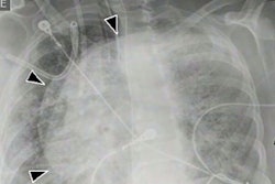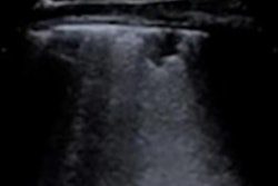
Lung ultrasound scans helped physicians in Europe manage the treatment of two patients with COVID-19 who required mechanical ventilation. The international team behind the cases published their report on September 9 in the American Journal of Tropical Medicine and Hygiene.
The physicians used lung ultrasound scores (LUS) to determine the severity of disease in two patients before and after starting mechanical ventilation. The scores helped the team to assess how well the patients responded to treatment in order to optimize their care.
"The two presented cases suggest the need for a personalized approach in use of adjunctive therapies for refractory hypoxemia," wrote the authors, led by Dr. Charalampos Pierrakos, an intensivist at Brugmann University Hospital in Brussels. "Lung ultrasound could be an attractive bedside tool to guide their use."
The two patients in the study were admitted to the hospital for laboratory-confirmed cases of COVID-19. The patients were in their 60s and obese, and both required invasive ventilation to manage refractory hypoxia. At admission, the CT scans of the patients showed ground-glass opacities involving 30% of the lungs.
The first patient, a 66-year-old woman, underwent invasive ventilation nine days after admission. Before starting ventilation, the authors calculated her lung ultrasound score. The LUS is the cumulative score from 12 individual sections of the lung. Each section can receive a score of 0-3, with higher scores indicating worse disease severity.
At the initial scan, the patient's cumulative LUS was 12, and none of her individual lung regions received the worst score of 3. But after the start of mechanical ventilation, the patient's total LUS shot up to 17. Several dorsal sites received a score of 3, indicating severe abnormal aeration.
Based on the score changes, the team performed an invasive recruitment maneuver by using continuous airway pressure at 60 cm H20 for 40 seconds. Following the maneuvers, the patient's LUS dropped to 14, mainly due to improvements in aeration of the dorsal areas. Her oxygen saturation also rose from 88% to 100%.
The team then moved the patient to a prone positioning. Her oxygenation and pulmonary system compliance both improved.
Similarly, the second patient, a 60-year-old man, underwent invasive ventilation 12 days after admission. An initial lung ultrasound scan showed an LUS of 11, and like the first patient, none of his lung regions had a score of 3.
For this patient, however, mechanical ventilation resulted in an improved LUS of 9. Based on the positive outcome, the team did not perform any recruitment maneuvers and left his ventilation settings the same. They also moved the patient to the prone position, and his oxygenation and pulmonary system compliance improved.
"These two cases illustrate how LUS could be useful in identifying different lung morphologies early after the start of invasive ventilation and help decide on adjunctive therapies," the authors wrote.
Lung ultrasound score has been studied by other researchers, with some findings suggesting that higher scores can indicate worse outcomes and even death from COVID-19. The new study adds to that research with further indication that ultrasound findings may also help to manage patient treatment.
Lung ultrasound also has other unique advantages for COVID-19, including that it can be used at bedside and be repeated as many times as needed. This is particularly important for assessing the immediate impacts of mechanical ventilation and potentially for performing resource management in places with limited resources.
"This has possible implications for ventilator management in resource-limited settings, with limited availability of chest computed tomography and blood gas analyzers," the authors wrote. "Tailoring invasive ventilation based on LUS findings early after the start of invasive ventilation is feasible, but this should be further evaluated in future studies."

![Examples of ultrasound findings and techniques. (A) Images in a 39-year-old male patient with a mass in the left thigh. The mass is heterogeneous on the B-mode US image (compared with the patient in D) and showed increased microvascularity (superb microvascular imaging [SMI]) and shear-wave elastography (SWE) values. Undifferentiated pleomorphic sarcoma was diagnosed at biopsy (with pleomorphic rhabdomyosarcoma in surgical specimen). (B) Images in an 18-year-old male patient with a mass in the left leg. The mass is hypoechoic on the B-mode image, with no other findings suggestive of malignancy. The lesion is in contact with the cortex of the tibia, which is slightly irregular. CT revealed a doubtful anteromedial tibial erosion. The microvascular study demonstrated high vascularization, suggestive of malignancy. Periosteal Ewing sarcoma was diagnosed with both histologic and immunohistochemical confirmation. (C) Images in a 69-year-old female patient with a lump growing on the outside of the left leg. Multiple SWE examinations were performed (please note the high values obtained in the measurements, whereas the color map highlights the stiffness relative to adjacent tissues). SMI showed areas of increased vascularization to target for sampling. Undifferentiated spindle cell sarcoma was diagnosed at biopsy, with residual leiomyosarcoma in the surgical specimen after neoadjuvant therapy. (D) Images in a 56-year-old female patient with a mass in the right thigh. The mass is heterogeneous at both B-mode ultrasound (similar to patient A) and MRI (coronal T2-weighted spectral attenuated inversion recovery [SPAIR]; T1-weighted pre-contrast and postcontrast imaging), which even shows uptake after the administration of paramagnetic contrast material, which is traditionally suggestive of malignancy. Low values at SMI and elastography are suggestive of benignity. Spindle cell lipoma was diagnosed at biopsy, with atypical spindle cell lipomatous tumor in the surgical specimen.](https://img.auntminnie.com/mindful/smg/workspaces/default/uploads/2026/08/images-radiol250278fig2.APCFLSvX6p.jpg?auto=format%2Ccompress&fit=crop&h=100&q=70&w=100)


![Examples of ultrasound findings and techniques. (A) Images in a 39-year-old male patient with a mass in the left thigh. The mass is heterogeneous on the B-mode US image (compared with the patient in D) and showed increased microvascularity (superb microvascular imaging [SMI]) and shear-wave elastography (SWE) values. Undifferentiated pleomorphic sarcoma was diagnosed at biopsy (with pleomorphic rhabdomyosarcoma in surgical specimen). (B) Images in an 18-year-old male patient with a mass in the left leg. The mass is hypoechoic on the B-mode image, with no other findings suggestive of malignancy. The lesion is in contact with the cortex of the tibia, which is slightly irregular. CT revealed a doubtful anteromedial tibial erosion. The microvascular study demonstrated high vascularization, suggestive of malignancy. Periosteal Ewing sarcoma was diagnosed with both histologic and immunohistochemical confirmation. (C) Images in a 69-year-old female patient with a lump growing on the outside of the left leg. Multiple SWE examinations were performed (please note the high values obtained in the measurements, whereas the color map highlights the stiffness relative to adjacent tissues). SMI showed areas of increased vascularization to target for sampling. Undifferentiated spindle cell sarcoma was diagnosed at biopsy, with residual leiomyosarcoma in the surgical specimen after neoadjuvant therapy. (D) Images in a 56-year-old female patient with a mass in the right thigh. The mass is heterogeneous at both B-mode ultrasound (similar to patient A) and MRI (coronal T2-weighted spectral attenuated inversion recovery [SPAIR]; T1-weighted pre-contrast and postcontrast imaging), which even shows uptake after the administration of paramagnetic contrast material, which is traditionally suggestive of malignancy. Low values at SMI and elastography are suggestive of benignity. Spindle cell lipoma was diagnosed at biopsy, with atypical spindle cell lipomatous tumor in the surgical specimen.](https://img.auntminnie.com/mindful/smg/workspaces/default/uploads/2026/08/images-radiol250278fig2.APCFLSvX6p.jpg?auto=format%2Ccompress&fit=crop&h=112&q=70&w=112)