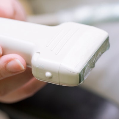
A group of radiology thought leaders is calling for a more precise definition of point-of-care ultrasound (POCUS). Their proposal to better distinguish POCUS from other ultrasound technologies was published on September 29 as a white paper in the Journal of the American College of Radiology.
The team behind the paper urged the medical community to see handheld ultrasound as the versatile innovation it is, noting some even liken its use to the stethoscope. But medical terminology and reimbursement codes have not kept up with the rapid and widespread advancement of ultrasound in medicine.
To remedy this, the Society of Radiologists in Ultrasound and the American College of Radiology Commission on Ultrasound came together to better delineate between observational and diagnostic ultrasound use of POCUS. In a new paper, the groups created four unique and distinct ultrasound categories, which include limiting the definition of POCUS to exams performed and reported in conjunction with a patient evaluation and management (E/M) encounter.
"We propose a simple way to broadly classify ultrasound evaluation that is more meaningful and consistent, taking the nomenclature beyond the idea of who performs the evaluation to the more important concept of what evaluation has been performed," wrote the authors, led by Dr. Maitray Patel, a radiologist at the Mayo Clinic Phoenix, AZ, and a member of both ultrasound societies behind the paper.
| 4 types of ultrasound evaluations: definitions and sample use cases | ||||
| Point-of-care ultrasound | Noncomprehensive diagnostic ultrasound | Partial-comprehensive diagnostic ultrasound | Complete-comprehensive diagnostic ultrasound | |
| Definition | Evaluation performed and reported in conjunction with an E/M encounter | Noncompre- hensive evaluation of any number of elements in anatomic region |
Comprehensive evaluation of a partial number of elements in an anatomic region | Comprehensive evaluation of a complete set of elements in an anatomic region |
| Example: pregnant patient in third trimester |
|
|
|
|
| Example: patient with right upper quadrant pain |
|
|
|
|
To create the four classifications, the authors first differentiated between point-of-care ultrasound (POCUS) and diagnostic ultrasound (DXUS) examinations. They asserted that POCUS becomes easier to recognize when defined solely as part of an E/M exam.
This definition of POCUS permits flexibility for what counts as a point-of-care exam. As described, POCUS would include scenarios that typically come to mind when thinking about point-of-care evaluations -- a clinician using a handheld device to scan a patient at bedside. But it also could include exams not obtained with handheld scanners or taken at bedside.
In addition to POCUS, the team created three distinct categories for diagnostic ultrasound use: noncomprehensive diagnostic ultrasound, partial-comprehensive diagnostic ultrasound, and complete-comprehensive diagnostic ultrasound.
The three diagnostic categories differ in relation to the author-defined principles of comprehensiveness and completeness. According to the group's terminology, comprehensiveness refers to the checklists and protocols needed to create, store, and document standardized images, whereas completeness recognizes whether the exam does or does not include all related structures or substructures in an anatomical region.
But this viewpoint is a major departure from how ultrasound is presently viewed in the larger medical infrastructure, particularly when it comes to billing and CPT codes.
"The current CPT framework forces the square POCUS peg exclusively through the round hole of 'diagnostic' imaging rather than considering and reimbursing POCUS as an inherent component or modifier of an E/M service," the authors wrote.
One of the challenges of the current reimbursement structure is that payors typically only pay for one documented and reported ultrasound evaluation of a patient, the authors noted. Instead, the new definition would permit multiple providers to perform POCUS evaluations on the same patient in the same region to answer multiple, relevant clinical questions.
In order to make the framework feasible, payers would have to recognize the benefits of allowing different providers to perform POCUS scans on the same patient. It would also require payors to work with multispeciality groups to define what organs and structures should comprise standardized anatomical subgroups.
"Make no mistake: There is important work to be done to understand the value of POCUS," the authors wrote.