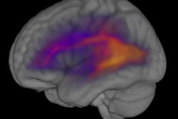
Functional MRI (fMRI) reveals that a brain region in the superior temporal sulcus (fSTS) is crucial for processing and making decisions about visual information and could provide information to treat visual conditions arising from stroke, according to a December 17 study in Neuron.
Researchers directly measured the firing of fSTS neurons in the areas of monkeys' brains as revealed by fMRI. The research team was led by Richard Krauzlis, PhD, chief of the National Eye Institute Section on Eye Movements and Selective Attention, which is part of the U.S. National Institutes of Health.
The monkeys fixed their eyes on a dot straight ahead and either paid attention to or specifically ignored stimuli happening in their visual periphery. The superior colliculus was strongly triggered when the monkeys paid attention to the visual event and a large proportion of fSTS neuronal activity was dependent on the superior colliculus.
When Krauzli and his team dampened the superior colliculus, the fSTS neurons showed less distinction between stimuli that the monkeys paid attention to versus what they ignored, meaning the fSTS depends on the superior colliculus to mark which stimuli are important and which are not.
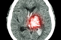
The findings are relevant for "visual neglect," which can occur after a stroke or other brain injury. People with visual neglect can see all the objects and events in their visual field, but they often aren't aware of the events on the affected side.
"Visual attention has to do with the internal management of information," Krauzlis said. "The connection with the superior colliculus is important because we think it could be acting like a spatial index that helps you keep track of the information that you're trying to process."