CAPE TOWN - Cardiovascular MR (CMR) and proton cardiac MR spectroscopy (¹H-cMRS) findings could serve as a cardiac imaging biomarker for at-risk women with HIV being treated via antiretroviral therapy (ART), according to research presented May 9 at the International Society for Magnetic Resonance in Medicine (ISMRM) annual meeting.
In her presentation, Petronella Samuels from the University of Cape Town in South Africa shared results showing how these imaging methods reveal myocardial steatosis and fibrosis in ART-treated women with HIV.
 Petronella Samuels presents her study at ISMRM 2026, which earned top honors for the category of Research Oral Presentations.
“We now see subtle diastolic dysfunction culminating in heart failure with preserved ejection fraction,” Samuels told AuntMinnie.com. “By using this data, we can see whether people are at risk of developing diastolic dysfunction and they can be treated earlier.”
Women with HIV have higher risk of developing heart failure, with the early era of ART believed to be a contributor. CMR and 1H-cMRS are the standard imaging modalities for detecting early myocardial tissue abnormalities linked to pre-heart failure, according to guidelines released in 2022 by the American Heart Association and the American College of Cardiology.
The Ugandan sTudy of HIV effects on the Myocardium and Atherosclerosis (MUTIMA) study was a prospective cohort study conducted between 2017 and 2019. It assessed the association of HIV with coronary artery disease.
In a sub-analysis of the MUTIMA study, Samuels and colleagues studied the prevalence of myocardial steatosis in ART-treated women with HIV in South Africa by using in vivo ¹H-cMRS.
The study included 17 ART-treated women living with HIV who had an average age of 53 years and average body mass index (BMI) of 30.8 kg/m2) and who had no known CVD. The researchers matched these women with 17 healthy HIV-negative women and used a 3T MRI scanner. After exclusions due to poor image quality, final analysis included 16 women with HIV and 11 controls.
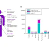
In women with HIV, CMR demonstrated normal biventricular functions and left ventricular mass index. These included the following data points: Left ventricular ejection fraction (LVEF), 63%; right ventricular ejection fraction (RVEF), 59%); and LV mass index, 41 g/m2.
Samuels also reported native T1, T2, and extracellular volume values being within normal ranges, including 1,233 ms, 40 ms, and 29%, respectively. These values did not point to diffuse myocardial fibrosis or edema.
However, 12 women with HIV had late gadolinium enhancement with mixed patterns, including the following: linear (n = 7), patchy (n = 2), diffuse (n = 2), and transmural (n = 1). These affected all areas of the myocardium, Samuels said.
Samuels also reported higher levels of intramyocardial triglyceride (IMTG) levels in five women with HIV, ranging from 1.4% to 3.1%.
Finally, three healthy controls had elevated IMTG levels, ranging from 0.5% to 1.1%. These corresponded to a higher average BMI of 30.9 kg/m2. However, imaging showed that their average IMTG values were within normal ranges.
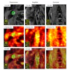
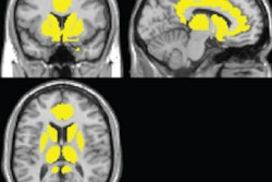
 Short-axis phase-sensitive-inversion-recovery (PSIR) sequences show different patterns of late gadolinium enhancement (LGE). A) Linear pattern; B) patchy; C) microvascular obstruction on early gadolinium enhancement (yellow arrow); and D) transmural LGE in patients with elevated IMTG levels.
Samuels said the results further reveal connections between HIV and CVD, as well as support the idea of metabolic and inflammatory myocardial remodeling.
She told AuntMinnie.com that the research is “a work in progress” and that data acquisition is still ongoing.
“We hope to have all the analysis in the next few months,” she said.
The research earned first place at ISMRM for the category of Research Oral Presentations.
“It’s a great opportunity. And for me, it’s extra special because Cape Town is my hometown,” she said of the award. “It’s a great honor to be recognized.”
Check out AuntMinnie.com’s full coverage of ISMRM 2026 on our ShowCast.