CAPE TOWN - MRI plays a key role in imaging athletes, especially during elite competitions such as the FIFA World Cup championships, according to a talk delivered May 9 at the ISMRM meeting.
Presenter Marcelo Bordalo, MD, PhD, of Aspetar Orthopedic and Sports Medicine Hospital in Doha, Qatar, offered session attendees a primer on how to provide imaging services to athletes during these competitions, using his experience at the FIFA World Cup event held in that country in 2022.
 Marcelo Bordalo, MD, PhDISMRM
"Why do we consider elite athletes different [from other patients]? Because their performance baseline isn't the same as the population norm, because we have to establish a return-to-play timeline, and because there are sport-specific microtrauma patterns," he said.
Bordalo's hospital provided imaging diagnosis via MRI, CT, x-ray, and ultrasound to 177 athletes during the competition -- with MRI the predominant modality. At the event, there were 832 athletes, 130 referees, and 1,300 FIFA staffers.
"Our goal was to make the teams feel as if they were in their own home countries in terms of medical imaging," he said.
One way the department did this was by establishing a protocol under which athletes from different teams did not encounter each other when they arrived for imaging. It also provided reports in English, Portuguese, Spanish, French, and Arabic and discussed all exams with the athletes' physician or physiotherapist. Finally, for all MRI exams, the department provided an audiovisual report, which improved evaluation time by 72%, Bordalo said.
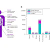
Of the total number of athletes, 143 imaging studies were conducted, for a use rate of 17.1%; of these, 67.7% were MRI exams, with digital x-ray coming in second at 11.8%. The majority of injuries were to the thigh, the knee, or the foot (27.2%, 23.2%, and 23.2%, respectively).
Bartolo offered the following "protocol pearls" for imaging elite athletes:
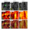
- Default to 3-tesla MRI, which provides higher signal-to-noise ratio for cartilage on T2/T2* imaging and better evaluation of small ligaments in the shoulder and elbow.
- Use multichannel surface coils: 16+ channel knee/shoulder coils allow for submillimeter, in-plane resolution.
- Use isotropic 3D to replace multiple, time-consuming 2D scans.
He also offered a framework for a variety of imaging scenarios, noting the importance of classifying muscle injuries for injury staging and prognosis, treatment planning (conservative versus surgical), and rehabilitation strategies and reinjury prevention). He listed five classification tools: Modified Peetrons, Chan, the British Athletics Muscle Injury Classification (BAMIC), Munich, and Barcelona/Aspetar, noting that BAMIC in particular is anatomically detailed and provides prognostic information.
Finally, Bordalo underscored three specific protocols, one for cartilage imaging (T2/T1 mapping); another for nerves (3D isotropic steady state sequences); a third for fatty infiltration grading (Dixon); and a fourth for groin imaging (using specific sequences rather than defaulting to a pelvic MRI exam).
In any case, Bordalo concluded that "MRI is the most used imaging modality for elite athletes."
Check out AuntMinnie.com’s full coverage of ISMRM 2026 on our ShowCast.