(Radiology Review) German radiologists recently compared the diagnostic accuracy of whole-body 3D contrast-enhanced magnetic resonance angiography (MRA) with digital subtraction angiography (DSA) in patients with peripheral arterial disease.
Researchers from the University Hospital Essen in Germany published their report in the American Journal of Roentgenology.
According to Dr Christoph Herborn and colleagues, the traditional diagnostic approach for atherosclerosis has been segmental because the risks associated with angiography have discouraged whole-body imaging.
According to the authors, MRA with 3D and contrast techniques provide a non-invasive method of assessing the presence and extent of atherosclerosis without the use of non-nephrotoxic contrast agents and ionizing radiation.
"The use of bolus chase techniques after a single injection of contrast medium now allows successive extended coverage to encompass vascular territories including the pelvic, femoral, popliteal, and calf arteries," they wrote.
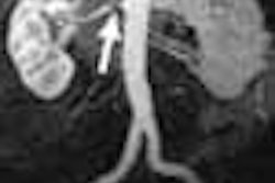
Using a table specifically designed for whole-body MA, the AngioSURF, six 3D data sets were acquired in succession. Selection of the field of view was achieved by adjusting stepping blocks and markers under the AngioSURF table. Using a sliding surface coil instead of a body coil resulted in better image quality.
Fifty-one patients with peripheral arterial occlusive disease referred to the hospital Essen for DSA also had whole-body MRA. Using a 1.5-T MR scanner, "paramagnetic gadobutrol was administered and five contiguous stations were acquired with 3D T1-weighted gradient-echo sequences in a total scanning time of 72 seconds."
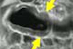
DSA results were considered the gold standard for assessing peripheral arterial disease. When new vascular disease was detected by whole-body MRA, the patients were further assessed using Doppler sonography, targeted MR angiography, or both.
"AngioSURF-based whole-body MR angiography had overall sensitivities of 92.3% and 93.1%, with specificities of 89.2% and 87.6% and excellent interobserver agreement (0.82) for the detection of high-grade stenoses," they reported.
Further assessment with whole-body MRA found additional atherosclerosis in 23% of patients.
Overall, they noted, "whole-body MR angiography permits a rapid, noninvasive, and accurate evaluation of the lower peripheral arterial system in patients with peripheral arterial occlusive disease, and it may allow identification of additional relevant vascular disease that was previously undetected."
AngioSURF provided highly accurate information in patients with peripheral arterial disease. This technique is capable of identifying unsuspected atherosclerosis and is ideal for diagnosing the extent of disease in patients with peripheral arterial disease, the group concluded.
Whole-Body 3D MR Angiography of Patients with Peripheral Arterial Occlusive DiseaseChristoph Herborn, et. al.
Department of diagnostic and interventional radiology, University Hospital Essen, Germany.
AJR 2004; 182:1427-1434
By Radiology Review
July 16, 2004
Copyright © 2004 AuntMinnie.com


![Overview of the study design. (A) The fully automated deep learning framework was developed to estimate body composition (BC) (defined as subcutaneous adipose tissue [SAT] in liters; visceral adipose tissue [VAT] in liters; skeletal muscle [SM] in liters; SM fat fraction [SMFF] as a percentage; and intramuscular adipose tissue [IMAT] in deciliters) from MRI. The fully automated framework comprised one model (model 1) to quantify different BC measures (SAT, VAT, SM, SMFF, and IMAT) as three-dimensional (3D) measures from whole-body MRI scans. The second model (model 2) was trained to identify standardized anatomic landmarks along the craniocaudal body axis (z coordinate field), which allowed for subdividing the whole-body measures into different subregions typically examined on clinical routine MRI scans (chest, abdomen, and pelvis). (B) BC was quantified from whole-body MRI in over 66,000 individuals from two large population-based cohort studies, the UK Biobank (UKB) (36,317 individuals) and the German National Cohort (NAKO) (30,291 individuals). Bar graphs show age distribution by sex and cohort. BMI = body mass index. (C) After the performance assessment of the fully automated framework, the change in BC measures, distributions, and profiles across age decades were investigated. Age-, sex-, and height-adjusted body composition reference curves were calculated and made publicly available in a web-based z-score calculator (https://circ-ml.github.io).](https://img.auntminnie.com/mindful/smg/workspaces/default/uploads/2026/05/body-comp.XgAjTfPj1W.jpg?auto=format%2Ccompress&fit=crop&h=100&q=70&w=100)


![Overview of the study design. (A) The fully automated deep learning framework was developed to estimate body composition (BC) (defined as subcutaneous adipose tissue [SAT] in liters; visceral adipose tissue [VAT] in liters; skeletal muscle [SM] in liters; SM fat fraction [SMFF] as a percentage; and intramuscular adipose tissue [IMAT] in deciliters) from MRI. The fully automated framework comprised one model (model 1) to quantify different BC measures (SAT, VAT, SM, SMFF, and IMAT) as three-dimensional (3D) measures from whole-body MRI scans. The second model (model 2) was trained to identify standardized anatomic landmarks along the craniocaudal body axis (z coordinate field), which allowed for subdividing the whole-body measures into different subregions typically examined on clinical routine MRI scans (chest, abdomen, and pelvis). (B) BC was quantified from whole-body MRI in over 66,000 individuals from two large population-based cohort studies, the UK Biobank (UKB) (36,317 individuals) and the German National Cohort (NAKO) (30,291 individuals). Bar graphs show age distribution by sex and cohort. BMI = body mass index. (C) After the performance assessment of the fully automated framework, the change in BC measures, distributions, and profiles across age decades were investigated. Age-, sex-, and height-adjusted body composition reference curves were calculated and made publicly available in a web-based z-score calculator (https://circ-ml.github.io).](https://img.auntminnie.com/mindful/smg/workspaces/default/uploads/2026/05/body-comp.XgAjTfPj1W.jpg?auto=format%2Ccompress&fit=crop&h=112&q=70&w=112)


