Hepatobiliary-phase hypointensity showed high interreader agreement among radiologists evaluating liver observations with LI-RADS CT/MRI (v2018) ancillary features (AFs) -- particularly those suggesting malignancy, researchers have found.
The results could have direct implications for how LI-RADS CT/MRI (v2018) should be applied in clinical liver imaging practice, according to a team led by Yong Jun Jung, MD, of the University of Ulsan College of Medicine in Seoul, South Korea. The study findings were published April 22 in the American Journal of Roentgenology.
"Interreader agreement was generally higher for AFs favoring malignancy than for those favoring benignity," the group noted.
In LI-RADS CT/MRI (v2018), AFs support observation characterization and category assignment, it explained. Unfortunately, there isn't much research regarding interreader agreement across the range of AFs.
To address this gap, Jung and colleagues conducted a review and meta-analysis of interreader agreement of all individual AFs in LI-RADS CT/MRI (v2018), identifying factors associated with interpretation variability. They searched Medline, Embase, and Cochrane databases for original research studies published between January 2018 and September 2025 that reported interreader agreement in these LI-RADS frameworks. Final analysis included 47 studies that represented 11,891 patients and 13,695 hepatic observations.
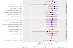
The team found that AFs favoring malignancy had higher interreader agreement than those that appeared benign:
Interreader agreement for individual ancillary features in LI-RADS CT/MRI (v2018) | |
Ancillary features (AFs) by type | Pooled kappa value |
AFs favoring malignancy but not HCC | |
| Hepatobiliary-phase hypointensity | 0.84 |
| Mild–moderate T2 hyperintensity | 0.77 |
| Restricted diffusion | 0.75 |
| Iron sparing in solid mass | 0.72 |
| Transitional-phase hypointensity | 0.70 |
| Fat sparing in solid mass | 0.69 |
| Corona enhancement | 0.63 |
| Subthreshold growth | 0.58 |
AFs favoring HCC | |
| Blood products in mass | 0.75 |
| Fat in mass more than adjacent liver | 0.72 |
| Mosaic architecture | 0.69 |
| Nodule-in-nodule appearance | 0.66 |
| Nonenhancing capsule | 0.58 |
AFs favoring benignity | |
| Iron in mass more than liver | 0.73 |
| Undistorted vessels | 0.60 |
| Marked T2 hyperintensity | 0.59 |
| Hepatobiliary-phase isointensity | 0.58 |
| Enhancement paralleling blood pool | 0.26 |
The finding that AFs suggesting benignity showed markedly lower agreement could have implications for how in LI-RADS CT/MRI (v2018) should be applied in clinical liver imaging practice, the group suggested, noting that the lower reproducibility of benignity-favoring features likely reflects their rarity in clinical practice, as they appear in only 1% to 3% of cases.
"Cautious application of AFs in clinical practice is advised for the AFs associated with low agreement, particularly in the settings further contributing to reduced agreement," the team concluded.
Access the full review here.