MONTREAL - Getting the contrast agent, dose, and timing right for the imaging task at hand remains one of radiology’s most challenging jobs. In two studies presented on Sunday at the International Congress of Radiology, European researchers sought to answer some interesting contrast questions for abdominal applications in both CT and MRI.
In an MDCT study of the abdomen, Dr. R. Hammersting from the University of Frankfurt am Mein in Germany, discussed her group’s study of Iomeprol (Bracco, Milan, Italy) -- administered in three different concentrations while maintaining the same overall iodine dose -- with the goal of determining which concentration offered the best lesion conspicuity.
And in an MRA study of the renal arteries, Dr. Mathias Prokop from the University of Utrecht in the Netherlands, working with colleagues throughout Europe, compared a single dose of Gd-BOPTA (Bracco) to a double dose of Gd-DTPA (Schering, Berlin, Germany).
Renal MRA
"T1 relaxivity in the blood is approximately twice as high for Gd-BOPTA (9.7 mmol)
as for Gd-DTPA (4.9 mmol); what that actually means is for the same amount of contrast you should be able to reduce the dose of Gd-BOPTA without reducing image quality," Prokop said. "We tried to compare the two techniques for contrast-enhanced MRA of the renal arteries."
The phase II crossover comparison study was conducted at the University of Vienna in Austria, at University Hospital in Essen, Germany, and at Hôpital Louis Pradel in Lyon, France.
Each patient underwent two MRA exams in random order: one with a single dose (0.1 mmol/kg) of Gd-BOPTA and the other with a double dose (0.2 mmol/kg) of Gd-DTPA. The second exam was always separated from the first by more than 48 hours but less than 12 days, Prokop said. Forty-one patients were included from the study, but six dropped out (three from each contrast group) before the second exam, leaving for comparison 34 patients who underwent exams with both agents.
After each contrast injection, breath-hold phase-encoded 3D-spoiled GRE images were acquired on 1.5-tesla MRI scanner, either a Magnetom Vision or a Symphony Sonata (Siemens Medical Solutions, Erlangen Germany), using TR 5 msec, TE 1.8 msec, flip angle 25° - 45°, FOV = 338-500 x 350-500 mm, with a minimum of 28 partitions, and slice thickness ranging from 1.0-3.3 mm.
"We used a test bolus injection to determine the start delay, and the flow rate was 2 ml per second," he said. "The evaluation was based on subracted MIP image, and of course in a random viewing order to avoid viewing biases."
The readers, blinded to all results, graded each of nine vessel segments on a scale of 0 to 2, (2 being adequate), with a possible overall score between 0 and 18. For quantitative assessment, vessel signal-to-noise ratio (SNR) and vessel-muscle contrast-to-noise ratio (CNR) were evaluated in supra-, juxta-, and infrarenal aorta psoas muscle.
"What we found … was no significant difference in the score of diagnostic information," Prokop said (reader 1: 15.15 and 15.23 for Gd-BOPTA and Gd-DTPA, respectively; reader 2: 16.77 and 17.01 for Gd-BOPTA and Gd-DTPA, respectively). The order of the exam had no significant effect, nor were there any differences in the mean SNR and CNR, though both tended to increase while descending the aorta. For the supra/juxta/infrarenal aorta respectively, the Gd-DTPA SNR was 38/37/41, while the CNR was 34/34/36, respectively. For Gd-BOPTA in the same three regions of interest, SNR was 32/42/48, and CNR was 34/38/44, he said.
 |
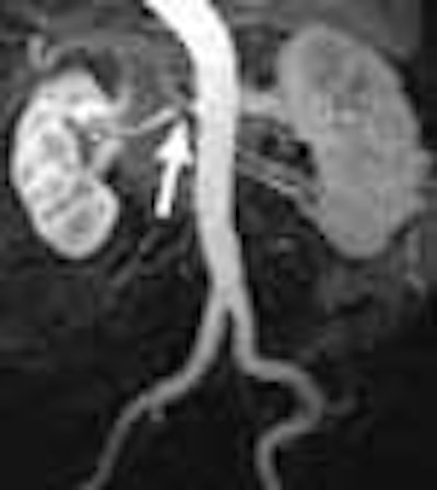
| Despite some unavoidable variations in contrast delivery timing that affect the image display, contrast enhancement in MR angiography is equivalent using either 0.1 mmol/kg Gd-BOPTA or 0.2 mmol/kg of Gd-DTPA. Images courtesy of Dr. Mathias Prokop. |
"You can see that timing does play a role in these images, so despite the fact that they were using the same technique and the same bolus injection, (timing) is obviously not 100% reproducible, and may contribute to variability in the display," Prokop said.
The conclusion is simple, he said. "It doesn’t matter whether you use a 0.1 mmol/kg Gd-BOPTA or 0.2 mmol/kg of Gd-DTPA, the evaluation of the renal arteries using these techniques is equivalent."
Whether there is any cost advantage in using one preparation over the other depends entirely on where it is purchased, he said in response to a question. Prices for the agents vary "massively" between countries, he said, so the price differential can range from 50% to nothing at all.
MDCT of the abdomen
"Due to the shorter scan duration with multidetector CT we now have well-defined phases of scanning regarding perfusion and enhancement," said Hammersting from Frankfurt, discussing her study of contrast-enhanced MDCT of the abdomen. But in order to keep the faster scans from outrunning the contrast, a choice must be made to either increase the injection flow rate or use higher-concentration contrast media, she said.
"We were interested in optimizing the iodine concentration in multidetector CT of the abdomen in order to (improve) therapeutic staging," Hammersting said.
The group performed a multicenter double-blinded stage IV randomized comparison of 91 patients for the evaluation of abdominal tumors. Sixty patients had liver cancer, and the rest, pancreatic, or gastric carcinoma. All of the patients underwent four-slice MDCT using a biphasic contrast-enhanced technique.
Before scanning, the researchers administered Iomeprol, a nonionic, monomeric contrast medium, in three different concentrations: 300, 350, 400 ml iodine/ml using an automatic power injector, while keeping the overall iodine dose constant at 36 g. Group 1 received 120 ml Iomeprol 300; group 2 received 105 ml Iomeprol 350; group 3 received 90 ml Iomeprol 400. The injection rate for all three groups was 4 ml/sec for the arterial phase, and 2 ml/sec for the portal-venous phase. Contrast timing was adjusted to the cardiac output of the patient.
"For our statistical analysis we used the absolute contrast between the lesion and the surrounding tissue, as well as contrast density for several organs," Hammersting said.
According to the results, contrast enhancement trended higher with the more concentrated contrast. For normal liver tissue in the arterial phase, contrast density was 61.3, 64.8, and 72.7 for Iomeprol 300, 350, and 400, respectively (p=0.01).
In addition to higher contrast in normal liver tissue, Iomeprol 400 produced slightly higher median contrast density in tissue surrounding lesions (74 HU versus 64, 66 for Iomeprol 300 and 350, respectively) and in the portal-venous phase (133 HU versus 99 and 102 for Iomeprol 300 and 350, respectively).
However, none of these differences were statistically significant. Statistical significance was reached in certain types of lesions, for which improved contrast density with Iomeprol 400 was marked: (71 HU versus 45 and 48 in primary liver carcinoma, and 84 versus 64 and 65 for cholangiocarcinoma). There was little improvement in contrast density in the arterial structures or pancreatic tissues with higher concentrations of iodine, Hammersting said. There were no adverse reactions in any of the patients.
The use of higher iodine concentrations while maintaining a constant iodine dose can provide better delineation and demarcation of some abdominal tumors in MDCT, Hammersting concluded.
By Eric Barnes
AuntMinnie.com staff writer
June 28, 2004
Copyright © 2004 AuntMinnie.com