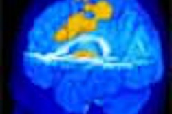
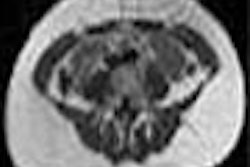
Combidex, an investigational MRI iron oxide nanoparticle contrast agent manufactured by Advanced Magnetics of Cambridge, MA, has shown encouraging results in the non-invasive diagnosis of metastatic lymph nodes. In two presentations at the International Society for Magnetic Resonance in Medicine, the product was determined to be a useful tool in characterizing cancerous lymph nodes.
The first presentation, by Dr. Mukesh Harisinghani, an assistant radiologist at Massachusetts General Hospital in Boston, offered data on the use of the contrast agent for characterizing lymph nodes in patients with breast cancer. Harisinghani found that the product had advantages in assisting physicians in staging cancers.
The second presentation, by Dr. Ralph Weissleder, director of the Center for Molecular Imaging Research at Massachusetts General Hospital, concluded that Combidex is a useful MRI contrast agent for characterizing lymph nodes in patients with prostate cancer, and that Combidex-enhanced images alone may suffice for lymph node characterization.
The contrast agent is the lead product in the firm’s development pipeline, and has received an approvable letter, subject to certain conditions, from the U.S. Food and Drug Administration for use in the diagnosis of metastatic lymph nodes. Advanced Magnetics is continuing to work with the FDA to resolve the outstanding issues from the approvable letter in an effort to bring Combidex to the market, according to the company.
By AuntMinnie.com staff writersJuly 15, 2003
Related Reading
Advanced Magnetics sets $10 million stock sale, July 3, 2003
Advanced Magnetics falls into red in Q1, January 16, 2003
MRI agent detects lymph node disease in prostate and renal cancers, December 4, 2002
Advanced Magnetics revenues decline, November 25, 2002
NCI to study Advanced Magnetics agents, October 1, 2002
Copyright © 2003 AuntMinnie.com