The Society of Nuclear Medicine and Molecular Imaging (SNMMI) highlighted a study showing that a novel quantitative PET- and MRI-based approach can objectively identify a recently recognized type of dementia.
Called limbic-predominant age-related TDP-43 encephalopathy, or LATE, the condition is often mistaken for Alzheimer’s disease, as it can currently only be definitively distinguished from Alzheimer's disease through postmortem neuropathology, the SNMMI said in a news release.
“This research may enable earlier differentiation of LATE and Alzheimer’s disease, or their co-existence in the clinic, guiding targeted diagnostic workup and personalized care for dementia patients,” the organization noted.
In the study, researchers created PET templates from autopsy-confirmed cases for LATE and Alzheimer's disease and then used z-score maps generated from the templates to analyze 944 F-18 FDG-PET scans from cases referred by cognitive disorder clinics. They then compared the clinical and quantitative MRI volumetry data across the groups.
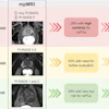
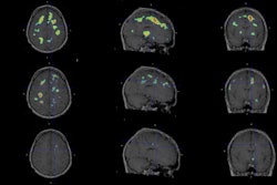
 Comparison of FDG-PET 3D-SSP z-score maps and transaxial slices of the temporal lobe, along with corresponding MRI images, among individuals suspected of having Alzheimer's disease, LATE, and mixed LATE and AD co-pathology. Color-coded areas on the 3D-SSP maps represent more severe metabolic reductions. Patients with LATE and LATE+AD show a distinct pattern of decreased FDG uptake in the medial temporal lobe, whereas AD and LATE+AD show the well-known pattern of decreased FDG uptake in the posterior cingulate cortex, precuneus, and parieto-temporal association cortices.Journal of Nuclear Medicine
"The imaging patterns identified on PET and MRI in this study provide clinicians with a practical tool to detect potential LATE pathology in patients with cognitive impairment and to inform clinical management and future investigations of LATE," wrote lead author Pei Ing Ngam, of the University of Utah in Salt Lake City, and colleagues.
The full study was published recently in the Journal of Nuclear Medicine and is available here.