A PET radiotracer for diagnosing Alzheimer’s disease may also be used to measure vascular brain changes in patients during PET/MRI scans, according to a study published December 7 in the Journal of Nuclear Medicine.
The finding suggests that hybrid PET/MRI can provide a more comprehensive picture of brain pathology involved in cognitive decline, noted first author Ates Fettahoglu, an undergraduate researcher at Stanford University in Stanford, CA, and colleagues.
“Two important biomarkers may be measured in a single session in participants with memory concerns, enabling improved characterization of dementia pathophysiology with reduced cost and inconvenience to participants and their caregivers,” the group wrote.
Both amyloid plaque brain deposits and changes in cerebral blood flow (CBF) are key components of Alzheimer’s disease, with amyloid PET scans and MRI typically used separately to detect changes in these biomarkers in patients.
While PET imaging with oxygen-15 (O-15) water radiotracer is the gold standard in molecular imaging for quantifying CBF, the use of this tracer is limited mostly to research settings due to its short half-life of two minutes, according to the researchers.
Moreover, previous work has suggested that “relative” CBF (flow between brain regions) can be measured with early-frame PET using F-18 amyloid tracers with half-lives of 110 minutes, the group noted. Still, obtaining measurement in absolute units (exactly how much blood is flowing) using this approach remains a challenge, they added.
To that end, the group hypothesized that they could accurately quantify CBF by combining early-frame F-18 florbetaben (Neuraceq, Life Molecular Imaging) scans with phase-contrast MRI scans of total brain blood flow during PET/MRI. This is also the first comparison of the use of F-18 florbetaben versus O-15 water PET to measure CBF, they added.
The researchers enrolled 20 participants. Eight patients were cognitively normal, nine had mild cognitive impairment, and three had dementia. Ten patients were negative for beta-amyloid plaque and 10 were beta-amyloid positive.
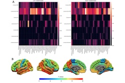
 A visual abstract of a study exploring early-frame F-18 florbetaben PET/MRI for quantifying cerebral blood flow in patients with cognitive impairment. Image courtesy of the Journal of Nuclear Medicine.
All patients underwent both O-15 water PET and phase-contract MRI and F-18 florbetaben and phase-contract MRI imaging in a single session on a 3-tesla PET/MRI scanner. The researchers quantified CBF by reconstructing images from the first two minutes of brain activity after radiotracer injections fused with phase-contrast MRI measurement of total brain blood flow.
A comparative analysis between the two approaches in patients showed similar results in gray matter blood flow, with a mean difference 0.7 milliliters per 100 grams of brain tissue per minute (0.7 ± 2.4 mL/100 g/min), according to the findings.
In addition, there were no significant differences in measuring CBF between the methods in the amyloid-positive and amyloid-negative groups as well as participants with different cognitive statuses, the authors found.
Ultimately, while amyloid PET scans are effective for diagnosing people with Alzheimer’s disease, some patients with beta-amyloid deposits are cognitively normal, the researchers noted. This suggests that a combined approach that also detects biomarkers such as changes in CBF can provide a more comprehensive picture, they wrote.
“[Early-phase F-18 florbetaben] PET/MRI can provide robust CBF measurements, highlighting the capability of simultaneous PET/MRI to provide measurements of both CBF and amyloid burden in a single imaging session in participants with memory disorders,” the group concluded.
A link to the full study is available here.