A deep-learning algorithm boosts CT imaging's performance when it comes to assessing patients with idiopathic pulmonary fibrosis, according to research presented at the RSNA meeting in Chicago.
The study results could translate to better patient outcomes, wrote a team of researchers led by presenter Dr. Ju Gang Nam of Seoul National University College of Medicine in South Korea.
"Predicting prognosis is important in determining treatment options and follow-up intervals in patients with idiopathic pulmonary fibrosis," Nam and colleagues noted. "Automatic CT quantification using deep learning may provide additional prognostic information in routine practice."
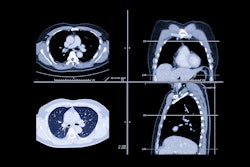
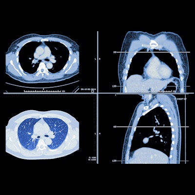
Idiopathic pulmonary fibrosis is a common interstitial pneumonia and has a high mortality rate. That's why being able to accurately assess the disease is key to effective patient management, Nam told attendees. CT plays an important role in the diagnosis of this condition and treatment assessment, but the degree of fibrosis isn't easily quantified on these exams, Nam explained.
"Radiologic prognostication of idiopathic pulmonary fibrosis is important, but [currently] there is no standard quantification method," he said.
Nam's group explored whether adding a deep-learning software to CT imaging could boost the modality's ability to quantify lung fibrosis in patients with the condition via a study that included 161 patients with diagnosed idiopathic pulmonary fibrosis who underwent noncontrast chest CT and spirometry between 2005 and 2009 within three months after receiving their diagnosis. The team used a commercially available deep-learning software (Aview by Coreline Soft) to determine proportions of normal lung volume (CT-Norm%) and fibrotic lung volume (CT-Fib%); they then correlated these results with spirometry results and calculated adjusted hazard ratios of CT-Norm% and CT-Fib%.
The researchers found that the combination of deep learning and CT imaging results showed that both CT-Norm% and CT-Fib% -- when adjusted for age, sex, smoking status, comorbidities of chronic diseases, and the spirometry measures of forced vital capacity (FVC), and diffusing capacity for carbon monoxide (DLCO) -- were predictors of patient survival.
"Normal lung and fibrotic lung volume proportions using a chest CT-based deep-learning algorithm ... correlated with physiologic variables and were independent predictors for overall survival in idiopathic pulmonary fibrosis," Nam concluded.