Deep-learning algorithms can effectively segment CT scans of patients with idiopathic pulmonary fibrosis (IPF) and thus offer prognostic information to clinicians, researchers have reported.
The data could be used to improve how patients with IPF are tracked and treated, wrote a team led by Munhunthan Thillai, MD, of the Royal Papworth Hospital in Cambridge, U.K. The group's findings were published August 14 in the American Journal of Respiratory and Critical Care Medicine.
"These segmentation algorithms could easily be integrated into routine clinical practice to help provide prognostic information to patients with this complicated and progressive lung disease, and they show promise as future key endpoints or imaging biomarkers in clinical trials," the researchers noted.
IPF is a disease that affects the tissue surrounding the air sacs in the lungs; it develops when that lung tissue becomes thick and stiff -- often without clear cause -- and over time, can lead to fibrosis, making it increasingly difficult for an individual to breathe, Thillai and colleagues explained. Previous studies have suggested that CT scans have a prognostic role in IPF based on image-based biomarkers, but these markers aren't regularly referred to in clinical practice or trials.
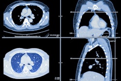
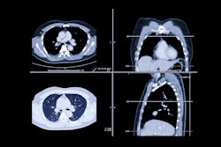
The authors tested the use of biomarkers (airway, lung, vascular, and fibrosis volumes) for diagnosis of IPF using deep learning-based segmentation of CT scans and applied them to data from a group of 446 not-yet-treated patients with IPF enrolled in the PROFILE (Prospective Observation of Fibrosis in the Lung Clinical Endpoints) study. They evaluated any relationship between the biomarkers and lung function, disease progression, and mortality. Median follow-up was 39.1 months and cumulative incidence of death was 277, or 62.1%, over five years after diagnosis of IPF.
The deep-learning algorithm successfully segmented 97.8% of the CT images, the team reported. It also found the following:
- Lung, vascular, and fibrosis volumes were associated with poorer five-year survival -- a result that persisted after the researchers adjusted for gender and age.
- Decreasing lung volume (hazard ratio [HR], 3.41, with 1 as reference; p = 0.009) and increasing fibrosis volume (HR, 2.23 p = 0.009) were associated with poorer survival.
- Lower lung volume (HR 0.98; p = 0.001), increased vascular volume (HR, 1.3; p = 0.001), and increased fibrosis volume (HR, 1.17; p < 0.001) were associated with reduced two-year progression-free survival.
The findings could translate to better monitoring of patients with IPF, according to the team.
"We demonstrate that CT scans from patients with IPF can be used to train and develop models that can rapidly segment CT scans at scale to produce data on fibrosis, vessel, airway, and lung volumes and that these can predict both progressive disease and mortality," the group concluded.
The complete study can be found here.