LOS ANGELES -- A novel PET radiotracer that can accurately detect deep vein thrombosis in the legs and reveal whether clots have migrated to the lungs has won this year’s Image of the Year award at the Society of Nuclear Medicine and Molecular Imaging (SNMMI) annual meeting.
The image is from a phase II trial and shows how F-18 GP1 PET/CT revealed multiple blood clots in the deep veins of the left leg (from the thigh to the calf) of a 75-year-old woman, as well as several clots in the right calf.
 (Left) F-18 GP1 PET/CT images from a 75-year-old woman show multiple blood clots in the deep veins of the left leg (from the thigh to the calf), as well as several clots in the right calf. Venous ultrasound confirms blood clots in left thigh, knee and calf veins. PET/CT also detects clots in both lungs, which are confirmed by contrast-enhanced CT image. (Right) 18F-GP1 PET/CT images from a 70-year-old woman show widespread blood clots throughout the body. In addition to clots in the deep veins of both legs and the arteries of both lungs (A), the scan also detected unexpected clots in several other areas, including blood vessels near the skull/head (B), spine (C), heart (D), and pelvis (E). Further evaluation revealed that the patient had antiphospholipid syndrome, an autoimmune condition that increases the risk of abnormal blood clot formation.Sangwon Han, MD, PhD, and SNMMI
In the study, a team from the University of Ulsan College of Medicine in Seoul, South Korea, assessed the diagnostic accuracy and tolerability of a F-18 GP1, a tracer 10 years in the making, lead researcher Sangwon Han, MD, PhD, Han told AuntMinnie.com. F-18 GP1 is derived from elarofiban and selectively binds to glycoprotein IIb/IIIa receptors on activated platelets.
“These findings suggest that a single whole-body PET scan could accurately evaluate clots in both the legs and lungs at the same time, potentially reducing the need for multiple tests while improving patient convenience,” he said.
Deep vein thrombosis (DVT) is a common disease in which blood clots form in the legs, and in some cases, travel to the lungs as a pulmonary embolism. It affects approximately 900,000 Americans a year according to the Centers for Disease Control and Prevention. Early detection is critical to ensure prompt treatment.
Between January 2020 and August 2025, Han and colleagues enrolled 46 consecutive patients between the ages of 19 and 79 years old who had a first episode of clinically suspected acute lower-extremity DVT. Within 14 days, patients underwent F-18 GP1 PET/CT scans.
On standard venous ultrasonography (VUS) -- performed within seven days of the PET/CT scans -- 22 patients were positive for proximal DVT and 24 were negative (six isolated distal DVT, 18 non-DVT). Compared, for proximal DVT, F-18 GP1 PET/CT demonstrated a sensitivity of 95% and a specificity of 92%. For distal DVT, the positive percent agreement between F-18 GP1 PET/CT and VUS was 96%, and the negative percent agreement was 90%.
Further, F-18 GP1 PET/CT identified pulmonary embolism in 22 patients, including 19 with proximal DVT, two with distal DVT, and one without DVT. Overall, F-18 GP1 PET/CT was well tolerated, with no drug-related adverse events, according to the research.
“This approach could serve as a platform technology for detecting clots throughout the body and even help to detect stroke or cardiovascular disease,” said SNMMI Scientific Program Committee Chair Giuseppe Esposito, MD, in a statement. “These images show just how powerful molecular imaging can be.”
Each year, SNMMI chooses an image that best exemplifies the most promising advances in the field of nuclear medicine and molecular imaging. This year, the SNMMI Image of the Year was chosen from nearly 1,500 abstracts submitted for the meeting.
Check out AuntMinnie’s full coverage of SNMMI 2026 on our ShowCast.

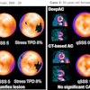


![(A-C) Representative whole-body maximum-intensity projection images and regional fused PET/CT images from three histologically confirmed osteosarcoma patients who underwent paired [68Ga]Ga-B7-H3-BCH PET/CT and 18F-FDGE PET/CT within seven days. (D) Multimodal imaging evaluation of Patient Three, including x-ray, MRI (T2-weighted imaging, T2WI), CT, and B7-H3 PET/CT.](https://img.auntminnie.com/mindful/smg/workspaces/default/uploads/2026/05/mei.XUQJWkpAJI.jpg?auto=format%2Ccompress&dpr=2&fit=crop&h=167&q=70&w=250)


![RET-targeted PET tracer highlights neuroendocrine prostate cancer tumors. Representative PET imaging shows strong tumor uptake of the RET-binding peptide tracer [⁶⁸Ga]Ga-DOTA-RET-L7 in a neuroendocrine prostate cancer (NEPC) model, supporting highly specific, high-contrast detection.](https://img.auntminnie.com/mindful/smg/workspaces/default/uploads/2026/05/screenshot-2026-05-27-205827.278Ys6PYU3.png?auto=format%2Ccompress&dpr=2&fit=crop&h=167&q=70&w=250)


