A portable, point-of-care PET technology can image organs and help guide interventional procedures, suggest findings presented June 1 at the 2026 Society of Nuclear Medicine and Molecular Imaging (SNMMI) annual meeting in Los Angeles.
In her talk, Xiyan Li, a graduate researcher from Washington University in St. Louis, described the success from the team’s PET technology, which proved to be workable in providing interactive PET scans and real-time image updating strategies.
“This proposed approach better supports interactive and adaptive imaging workflows at the bedside,” Li said.
Previous research suggests that PET/CT-guided interventional radiology procedures are more accurate than current interventional procedures that rely on anatomical imaging guidance from ultrasound, x-ray, fluoroscopy, and CT.
Li and colleagues suggested that their ordered-subset expectation-maximization (OSEM)–based reconstruction framework could make way for real-time image updates when scanning a patient from multiple angles interactively.
The researchers studied the feasibility of their point-of-care, interactive PET scanning system. They used a phantom containing three clusters of radiotracer-filled rods imaged by the system, moving the detector panels to six user-selected positions.
The team reconstructed imaging data beginning with five iterations from the first position. This was followed by alternating single-iteration updates through data taken from each new position. The researchers continuously updated images as they were acquired, as data acquisition time took “significantly” longer than reconstruction time. They also compared this approach to that of a conventional PET reconstruction framework based on maximum-likelihood expectation-maximization (MLEM).
Li reported that the team’s approach led to fine phantom structures becoming clearly distinguishable after four positions. He added that this result suggests that scanning could be ended early and image quality can be improved with additional OSEM iterations.
In addition, incrementally updated OSEM images proved to be comparable to conventional MLEM reconstructed images (20 iterations) using all six data sets. The OSEM images also provided continuous visual feedback to the operator during acquisition.
Each OSEM update loop needed about two seconds while 20-iteration MLEM reconstruction needed about nine seconds on an
For a total data size of 98.3 MB (16.3 MB in each subset of data), 20-iteration MLEM reconstruction required approximately nine seconds on an Nvidia GTX 1660 Ti graphics card. The team also highlighted the proposed framework’s ability to update images as soon as scanning starts. MLEM images have reconstruction performed only after full acquisition.
 (a) Benchtop portable POC-PET prototype system. (b) Phantom imaging experiment setup. (c) Comparison of conventional full-data MLEM reconstruction with incremental OSEM reconstruction.Xiyan Li and SNMMI
Finally, Li noted that total OSEM computation time increases with the number of subsets. However, he added that the delays per-update were insignificant compared to the four-minute acquisition time per position. This allows for continuous real-time image updates and visual feedback, Li said.
“It [OSEM approach] represents a paradigm shift that offers new avenues to deploy novel molecular imaging applications,” Li said.
The team next plans to focus their work on improving their data subset design and AI-guided update ordering. It will also perform quantitative evaluation using contrast recovery, spatial resolution, and image uniformity metrics. Finally, the researchers are building a prototype system for analyzing human patients, with this study planned to begin in 2027.
Check out AuntMinnie’s full coverage of SNMMI 2026 on our ShowCast.
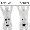


![(A-C) Representative whole-body maximum-intensity projection images and regional fused PET/CT images from three histologically confirmed osteosarcoma patients who underwent paired [68Ga]Ga-B7-H3-BCH PET/CT and 18F-FDGE PET/CT within seven days. (D) Multimodal imaging evaluation of Patient Three, including x-ray, MRI (T2-weighted imaging, T2WI), CT, and B7-H3 PET/CT.](https://img.auntminnie.com/mindful/smg/workspaces/default/uploads/2026/05/mei.XUQJWkpAJI.jpg?auto=format%2Ccompress&dpr=2&fit=crop&h=167&q=70&w=250)


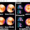
![RET-targeted PET tracer highlights neuroendocrine prostate cancer tumors. Representative PET imaging shows strong tumor uptake of the RET-binding peptide tracer [⁶⁸Ga]Ga-DOTA-RET-L7 in a neuroendocrine prostate cancer (NEPC) model, supporting highly specific, high-contrast detection.](https://img.auntminnie.com/mindful/smg/workspaces/default/uploads/2026/05/screenshot-2026-05-27-205827.278Ys6PYU3.png?auto=format%2Ccompress&dpr=2&fit=crop&h=167&q=70&w=250)


