LOS ANGELES -- A novel PET imaging approach enables simultaneous visualization of tumor progression and cardiac inflammatory responses during cancer treatment, according to research from the Society of Nuclear Medicine and Molecular Imaging (SNMMI) meeting.
A team at Washington University in St. Louis developed a novel radiotracer called copper-64 (Cu-64) DOTA-ECL1i that targets a protein called CCR2 (C-C motif chemokine receptor 2). CCR2 guides the migration of immune cells to sites of inflammation and is expressed in both atherosclerotic plaques and tumors, noted lead researcher Jaume Otaegui, MD.
“CCR2 PET imaging could potentially allow clinicians to detect harmful cardiovascular changes that occur during cancer treatment before they result in heart attack, myocarditis, or other serious complications,” Otaegui said, in a SNMMI statement.
Immune checkpoint inhibitor (ICI)-based therapies have revolutionized cancer treatment and substantially improved survival rates. Yet these treatments can also lead to serious heart and vascular adverse effects in a subset of patients. In this study, the group evaluated whether Cu-64 DOTA-ECL1i PET imaging could detect immune-related adverse events in a cardio‑oncology mouse model, as well as a therapeutic strategy to reduce cardiac risk during treatment.
The researchers implanted mouse oral carcinoma cells into two cohorts of atherosclerotic mice as a cardio-oncology model. The first cohort received ICI treatment or a control twice weekly, with Cu-64 DOTA-ECL1i PET performed after five and 10 doses; F-18 FDG PET/CT was also conducted in parallel for comparison. The second cohort received three weeks of ICI treatment, with itacitinib -- a Janus kinase 1 inhibitor -- added twice daily during the final two weeks, followed by Cu-64) DOTA-ECL1i PET imaging at the treatment endpoint.
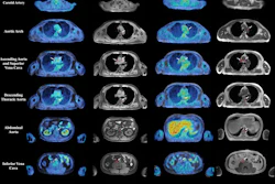
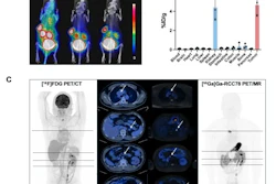
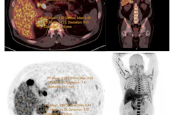
According to the results, Cu-64 DOTA-ECL1i demonstrated effective binding to CCR2-expressing cells and produced superior PET/CT visualization of both atherosclerotic plaque and tumor tissue compared with F-18 FDG PET. ICI treatment slowed down tumor progression, but did not affect CCR2 PET tumor signal. The CCR2 cardiac signal, however, was significantly increased. Cotreatment with itacitinib showed better tumor response, reduced CCR2 PET signal, and fewer inflammatory cells in the aorta compared to ICI treatment alone, the researchers reported.
 Combination therapy of JAK1i with anti-PD1 slows down tumor growth and reduces CCR2+ cells infiltration in atherosclerotic lesions compared to anti-PD1 alone. Representative PET images of cardio-oncology mice treated with anti-PD1 alone or in combination with JAK1i. The PET uptake was normalized by subtracting blood retention. Jaume Otaegui, MD, and SNMMI
“Our therapeutic approach of combining itacitinib ICI treatment could be used to both reduce cardiovascular inflammation and enhance tumor control.”
Ultimately, the study establishes a potential dual-purpose imaging and therapeutic strategy for cardio-oncology, Otaegui noted.
"Currently, there is no established molecular imaging approach to identify the development of these cardiovascular immune-related adverse events in patients receiving ICI treatments," he said.
Check out AuntMinnie’s full coverage of SNMMI 2026 on our ShowCast.