LOS ANGELES -- A novel tau radiotracer appears to successfully detect chronic traumatic encephalopathy (CTE)-associated tau pathology in living patients, according to research presented at the Society of Nuclear Medicine and Molecular Imaging (SNMMI) 2026 meeting.
The study findings could establish "a clinical role for PET in traumatic brain injury and sports- and military-related neurodegeneration and spark next generation tau-radiopharmaceuticals optimized for non-Alzheimer's disease tauopathies -- including CTE," lead researcher Isabelle Boileau, PhD, of the Centre for Addiction and Mental Health in Toronto, said in a statement released by the society.
"While this radiopharmaceutical is in the early clinical research stage, our data in both people with suspected CTE and other non-Alzheimer’s disease tauopathies is very promising," she noted.
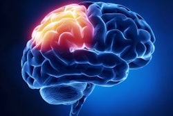
CTE is a trauma-associated tauopathy defined by clumps of abnormal tau protein that form around tiny blood vessels in the deep folds of the brain's outer surface -- affecting both cortical and white-matter regions. It is currently diagnosed postmortem, according to the researchers.
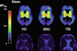
Boileau and colleagues developed a new pan-tau PET tracer called 18F-OXD-2314 to identify tau aggregates in both Alzheimer's and non-Alzheimer's tauopathies. They conducted a study that included seven healthy controls and three retired collision-sport athletes with presumed CTE, all of whom underwent a 120-min brain PET exam following and injection of approximately 190 MBq of 18F-OXD-2314. The study participants also underwent a brain MRI to identify regions of interest, including cortical grey ROIs (frontal, temporal, parietal, and occipital) and a gray–white matter junction region at the cortical sulci.
Key findings the researchers reported were that distribution volume ratio (DVR) was somewhat elevated in cortical regions in individuals with suspected CTE compared to healthy controls, significantly higher at the gray–white matter junction, and "robustly elevated" in whole-brain white matter.
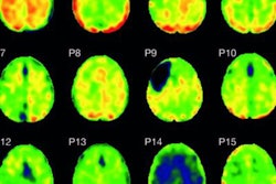
 Parametric maps obtained with SRTM2 (cerebellum grey matter as reference) for the group of healthy participants (top) and from one representative participant with suspected CTE (bottom). CTE: chronic traumatic encephalopathy. Isabelle Boileau, PhD, and SNMMI
This last finding is new, the team explained.
"The detection of activity in the white-matter is a novel observation on tau PET and may reflect trauma-related tau deposition along axons or within astrocytes and oligodendrocytes not detected by current tracers," it wrote.
If the study results can be validated, 18F-OXD-2314 could help provide "the first accurate in-life diagnostic biomarker for CTE," according to Boileau.
"While this radiopharmaceutical is in the early clinical research stage, our data in both people with suspected CTE and other non-Alzheimer's disease tauopathies is very promising," she said.
Check out AuntMinnie’s full coverage of SNMMI 2026 on our ShowCast.


![(A-C) Representative whole-body maximum-intensity projection images and regional fused PET/CT images from three histologically confirmed osteosarcoma patients who underwent paired [68Ga]Ga-B7-H3-BCH PET/CT and 18F-FDGE PET/CT within seven days. (D) Multimodal imaging evaluation of Patient Three, including x-ray, MRI (T2-weighted imaging, T2WI), CT, and B7-H3 PET/CT.](https://img.auntminnie.com/mindful/smg/workspaces/default/uploads/2026/05/mei.XUQJWkpAJI.jpg?auto=format%2Ccompress&dpr=2&fit=crop&h=167&q=70&w=250)


![RET-targeted PET tracer highlights neuroendocrine prostate cancer tumors. Representative PET imaging shows strong tumor uptake of the RET-binding peptide tracer [⁶⁸Ga]Ga-DOTA-RET-L7 in a neuroendocrine prostate cancer (NEPC) model, supporting highly specific, high-contrast detection.](https://img.auntminnie.com/mindful/smg/workspaces/default/uploads/2026/05/screenshot-2026-05-27-205827.278Ys6PYU3.png?auto=format%2Ccompress&dpr=2&fit=crop&h=167&q=70&w=250)


