

After a patient's face was burned from metal in a mask during a 3-tesla MRI scan, the U.S. Food and Drug Administration (FDA) on December 7 issued reminder guidance to patients and healthcare providers regarding wearing metal during MRI exams.
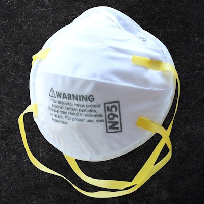
"Today, the [FDA] is informing patients and healthcare providers that patients may be injured if they wear face masks (such as surgical or nonsurgical masks and respirators) with metal parts and coatings during an MRI exam," the agency said. "The FDA reminds patients and providers that patients should not wear any metal during an MRI."
Because patients must wear masks to prevent the potential transmission of COVID-19, the issue has become urgent, the agency said in its statement. The patient who was injured underwent a 3-tesla MRI exam of the neck and was burned in a pattern consistent with the mask outline, the agency said.
"Given the increased use of face masks during the COVID-19 pandemic, the FDA wants patients and healthcare providers to be aware of the potential risk of face burns related to the use of patient face masks containing metal during an MRI," the agency said. "Metal parts ... [in a face mask] may become hot and burn the patient during an MRI."
It's appropriate for patients to wear face masks during an MRI exam due to the COVID-19 pandemic, the FDA said. But some face masks contain metal parts, like nose clips or wires, nanoparticles, or antimicrobial coating that may contain metal such as silver or copper. Before the MRI scan begins, healthcare providers who perform MRI exams should confirm the face mask has no metal.
"The metal could result in radio frequency (RF)-induced heating," the agency said. "This may represent a hazard for MR imaging during the COVID-19 pandemic."
The solution? Don't allow patients to wear their own masks during an MR exam, said Tobias Gilk, senior vice president of Radiology-Planning, founder of Gilk Radiology Consultants, and an MRI safety advocate.
"The staples holding the elastic to the mask are too small to conduct heat, and since the COVID-19 pandemic, patients have been imaged in masks that have nose bridges without injury," he said. "But antimicrobial treated fabric can heat up. So to be safe, patients should be provided with disposable surgical masks before their MRI."
The FDA asks any patients who are burned by a face mask during an MRI scan -- or providers who witness this kind of patient injury -- to report the event. It also refers patients and providers to American College of Radiology (ACR) resources regarding MRI exams during the COVID-19 pandemic.


![Overview of the study design. (A) The fully automated deep learning framework was developed to estimate body composition (BC) (defined as subcutaneous adipose tissue [SAT] in liters; visceral adipose tissue [VAT] in liters; skeletal muscle [SM] in liters; SM fat fraction [SMFF] as a percentage; and intramuscular adipose tissue [IMAT] in deciliters) from MRI. The fully automated framework comprised one model (model 1) to quantify different BC measures (SAT, VAT, SM, SMFF, and IMAT) as three-dimensional (3D) measures from whole-body MRI scans. The second model (model 2) was trained to identify standardized anatomic landmarks along the craniocaudal body axis (z coordinate field), which allowed for subdividing the whole-body measures into different subregions typically examined on clinical routine MRI scans (chest, abdomen, and pelvis). (B) BC was quantified from whole-body MRI in over 66,000 individuals from two large population-based cohort studies, the UK Biobank (UKB) (36,317 individuals) and the German National Cohort (NAKO) (30,291 individuals). Bar graphs show age distribution by sex and cohort. BMI = body mass index. (C) After the performance assessment of the fully automated framework, the change in BC measures, distributions, and profiles across age decades were investigated. Age-, sex-, and height-adjusted body composition reference curves were calculated and made publicly available in a web-based z-score calculator (https://circ-ml.github.io).](https://img.auntminnie.com/mindful/smg/workspaces/default/uploads/2026/05/body-comp.XgAjTfPj1W.jpg?auto=format%2Ccompress&fit=crop&h=100&q=70&w=100)


![Overview of the study design. (A) The fully automated deep learning framework was developed to estimate body composition (BC) (defined as subcutaneous adipose tissue [SAT] in liters; visceral adipose tissue [VAT] in liters; skeletal muscle [SM] in liters; SM fat fraction [SMFF] as a percentage; and intramuscular adipose tissue [IMAT] in deciliters) from MRI. The fully automated framework comprised one model (model 1) to quantify different BC measures (SAT, VAT, SM, SMFF, and IMAT) as three-dimensional (3D) measures from whole-body MRI scans. The second model (model 2) was trained to identify standardized anatomic landmarks along the craniocaudal body axis (z coordinate field), which allowed for subdividing the whole-body measures into different subregions typically examined on clinical routine MRI scans (chest, abdomen, and pelvis). (B) BC was quantified from whole-body MRI in over 66,000 individuals from two large population-based cohort studies, the UK Biobank (UKB) (36,317 individuals) and the German National Cohort (NAKO) (30,291 individuals). Bar graphs show age distribution by sex and cohort. BMI = body mass index. (C) After the performance assessment of the fully automated framework, the change in BC measures, distributions, and profiles across age decades were investigated. Age-, sex-, and height-adjusted body composition reference curves were calculated and made publicly available in a web-based z-score calculator (https://circ-ml.github.io).](https://img.auntminnie.com/mindful/smg/workspaces/default/uploads/2026/05/body-comp.XgAjTfPj1W.jpg?auto=format%2Ccompress&dpr=2&fit=crop&h=167&q=70&w=250)


