SALT LAKE CITY - The more disparate and dispersed a network is, the greater the need for it to be centrally managed. With the first wave of PACS implementation beginning to crest across the U.S., service and support of these networks is now coming to the forefront of information system (IS) managers’ concerns.
"We were tired of reacting to every PACS problem, instead of identifying and solving PACS/RIS issues before they became a problem," said Louis Lannum, an IS manager for the Cleveland Clinic Foundation (CCF) in Cleveland. He, and his 17-person IS team, made a commitment to prevent rather than put out PACS fires.
A multivendor suite of products now manages the CCF’s enterprise-wide PACS, said Lannum in a presentation on Thursday at the Symposium for Computer Applications in Radiology (SCAR) in Salt Lake City.
Lannum’s enterprise network covers a 100-square mile area in northeast Ohio and comprises 45 diagnostic radiology-viewing stations, more than 200 referring physician workstations, and multiple image storage systems on the main campus. Prior to centralizing the network management of the PACS, the radiology informatics group was divided into three service organizations.
A PACS group handled picture archiving and communication issues, a system group dealt with RIS and dictation systems, and a help desk fielded hardware and desktop user support. This Balkanization of responsibilities left the help-desk personnel constantly dealing with user-discovered problems. "A lot of people ended their day with a ‘Survivor’ mentality."
Lannum, determined to increase the level of support and improve the quality of service in the radiology division, turned to software application tools to monitor the mission-critical systems of the CCF PACS. He identified three operational goals he and his team sought to meet by centralizing the CCF PACS management.
First, the team wanted timely problem identification and resolution capabilities. Second, they wanted to increase system availability through monitoring the "health" of their network to identify potential trouble spots. Third, they wanted to be able to perform software installations and distribute software upgrades electronically over their system.
"Software upgrades that we used to be able to complete in a day or two, when our PACS was in one building, were now taking us up to several weeks to finish," Lannum said.
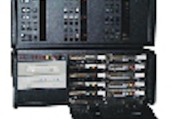
The CCF group implemented the MagicWatch (Siemens Medical Solutions, Iselin, NJ) application to track the status of every node on its PACS. The product, based on Palo
Alto, CA-based Hewlett-Packard’s Open View network-management software, electronically polls each switch, router, printer, and workstation on the PACS. The software is also capable of configuring the operating systems and PACS applications running on each workstation.
 |
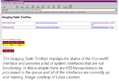
In addition to monitoring the PACS environment, the CCF team has put into place a centralized RIS-management application. The Imaging Suite Toolbox (IDX Systems, Burlington, VT) displays the system status of all interfaces in the RIS. This permits the IS team to assess traffic patterns across the RIS and alert users to potential delays in receiving images.
 |
Lannum reported that application monitoring is performed on a 16-hour schedule daily. He noted that with the continual monitoring of each node, the operations-center team has been able to identify potential hardware problems and notify the IS department before it becomes a user issue.
"There are a lot of system-monitoring tools on the market," Lannum said. "The important issue that IS administrators have to decide on is how they are going to use those tools."
By Jonathan S. Batchelor
AuntMinnie.com staff writer
May 3, 2001
Related Reading
Siemens emphasizes workflow at ECR, March 5, 2001
How far has systems integration evolved in PACS? February 20, 2001
Siemens Dashboard ready to drive, February 5, 2001
Stentor teams up with IDX, November 16, 2000
Kaiser Permanente to buy IDXrad for California sites, June 16, 2000
Click here to post your comments about this story in our PACS Digital Community. Please include the headline of the article in your message.
Copyright © 2001 AuntMinnie.com