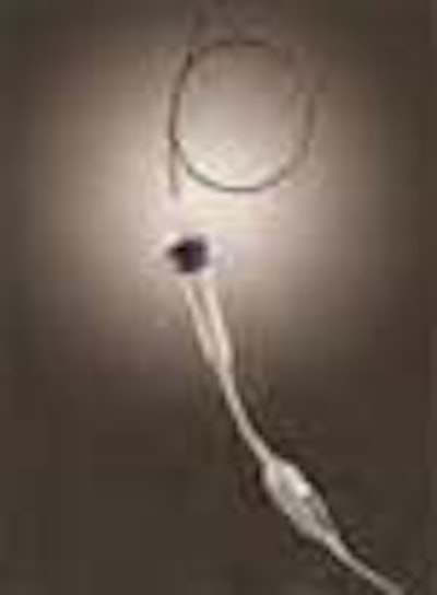
Acuson of Mountain View, CA, has announced that it has received 510(k) clearance from the U.S. Food & Drug Administration (FDA) to market the AcuNav diagnostic ultrasound catheter. The new tool brings the full performance of diagnostic ultrasound into the heart and may aid in the diagnosis and management of treatment heart disease and irregular heart rhythms, according to the company's press release.
The new intracardiac ultrasound technique works by inserting a catheter through the femoral vein of the leg or the jugular vein in the neck, then maneuvering it into the right atrium or right ventricle of the heart. The catheter's ability to image cardiac structures within the heart also shows potential to expand the utility of ultrasound into areas of electrophysiology, where ultrasound has previously played a limited role.
"This device enables us to clearly see areas of anatomy that govern heart function we have not been able to visualize previously," said James Seward, M.D., a Mayo Clinic cardiologist, director of Mayo Clinic's Echocardiography Laboratory in Rochester, Minn., and one of the inventors of the device. "This catheter with the eyes of ultrasound allows for more precise imaging within the heart and blood vessels."
Initial results of clinical trials indicate that cardiac structures are seen with diagnostic quality 100 percent of the time, according to the company. Prior to clearance, the AcuNav catheter was used for two emergency, life-saving procedures.
In the two isolated cases, physicians used the AcuNav catheter to help guide patient treatment when they were unable to properly visualize cardiac anatomy using available imaging technologies. One case at the University of Michigan Medical Center involved a patient suffering from end-stage primary pulmonary hypertension. The second case involved a patient with pulmonary vein stenosis.
Acuson has a license agreement with Mayo Clinic for use of the new tool. The Mayo Clinic and Acuson each hold a series of patents on, or relating to, the new technology, which was originally conceived by two Mayo Clinic cardiologists, Dr. Seward and A. Jamil Tajik, M.D., chair of Mayo Clinic's Division of Cardiovascular Diseases. A separate team led by Douglas Packer, M.D., a Mayo Clinic electrophysiologist, tested the tool's effectiveness in imaging cardiac anatomy and physiology, and other devices in the heart.
The company anticipates that shipments of the new catheter will begin in the first quarter of 2000.
By AuntMinnie.com staff writersDecember 21, 1999
Copyright © 1999 AuntMinnie.com






