Bronchiectasis:
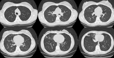
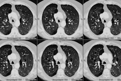
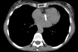
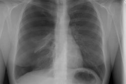
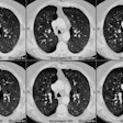
The patient shown in the CT scan below had a history of a severe respiratory infection for which she did not seek medical attention. The CT scan demonstrates extensive bronchiectatitc changes- more pronounced in the right lung- particularly in the right middle lobe. There is some nodular air space disease in the right middle lobe which may reflect mucous plugging of the distal airways. Other etiologies for extensive bronchiectasis such as cystic fibrosis, dysmotile cilia syndrome, and immunodeficiency syndromes should be excluded. Click here to view zoomed images of the upper and lower lobes.
