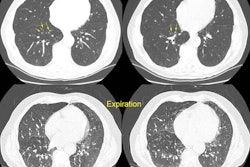
Bronchiectasis:
The CT scan demonstrates dilated, thick walled bronchi within the left lower lobe (red arrow). The distal bronchi are mucous filled, but should not be mistaken for vessels (yellow arrows). The density in the posterior right costophrenic sulcus was related to an area of atelectasis.