Lung Torsion
Clinical:
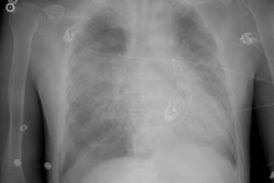
Lung torsion is a rare entity. Most cases invovle a single lobe [3] and develop after lobectomy because the surgical division of the pulmonary ligament encourages increased mobility of the remaining lobes. Torsion of the right middle lobe after a right upper lobectomy is the most frequently seen variantr [3]. Patients with complete fissures are at particularly increased risk and many surgeons affix the right middle lobe to the remaining lobe to prevent post-operative torsion. Torsion is unusual in the native lung and exceedingly rare after lung transplant. The degree of rotation in pulmonary torsion is generally 180 degrees [2]. Torsion compromises the three pulmonary vascular systems and the onset of gangrene is rapid with complete torsion. Most patients present with chest pain and hemoptysis. The mortality rate is high if the condition goes unrecognized [2].
X-ray:
The torsed lobe or lung often presents as a consolidated or collapsed lobe with an unusual position. CT will demonstrate bronchial obstruction and an abnormal arterial and venous relationships [2]. There will be tapered obliteration of the proximal pulmonary artery and accompanying bronchus. There is often an amorphous soft-tissue attenuation at the hilum [2]. The torsed lobe demonstrates poorly enhancing consolidation [2].
REFERENCES:
(1) AJR 2000; Gilkeson RC, et al. Lung torsion after lung transplantation: Evaluation with helical CT. 174: 1341-1343 (No abstract available)
(2) Radiographics 2002; Kim EA, et al. Radiographic and CT findings
in
complications following pulmonary resection. 22: 67-86
(3) Radiographics 2013; Lubner MG, et al. Emergent and nonemrgent nonbowel torsion: spectrum of imaging and clinical findgins. 33: 155-173