Radiology 1997 Apr;203(1):202-206. Telescoping bronchial anastomoses for unilateral or bilateral sequential lung transplantation: CT appearance.
McAdams HP, Murray JG, Erasmus JJ, Goodman PC, Tapson VF, Davis RD
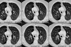
PURPOSE: To determine the computed tomographic (CT) appearance of telescoping bronchial anastomoses. MATERIALS AND METHODS: CT scans obtained in 36 adult patients with 54 telescoping anastomoses (30 right bronchus, 24 left bronchus) were retrospectively reviewed. Seventeen scans were obtained with 10-mm collimation, one with 5-mm collimation, and 18 with 3-mm collimation. Multiplanar volume reconstruction images were retrospectively generated in 19 patients. CT findings were correlated with fiberoptic bronchoscopic findings and clinical records. RESULTS: Smooth, spherical air collections caused by diverticula of redundant mucosa were seen at the inferior or medial aspect of 22 anastomoses (41%). Endoluminal flaps and linear air collections caused by separation of the invaginated bronchus from the recipient bronchus were seen in 16 anastomoses (30%). Flaps and linear air collections were seen only at the anterior, superior, or inferior aspects of the anastomosis. Flaps and diverticula were more common at right anastomoses than at left anastomoses. No patient with an endoluminal flap or diverticulum at CT had dehiscence at bronchoscopy. Irregular air collections or posterior wall defects were suggestive of dehiscence at four anastomoses (7%). Dehiscence was confirmed bronchoscopically at two of these four anastomoses. CONCLUSION: Endoluminal flaps and linear or spherical air collections can be a normal feature of the telescoping anastomosis and should not necessarily be interpreted as dehiscence.
PMID: 9122393, MUID: 97232423

