Asthma:
Clinical:
Asthma is a disease of the airways that is characterized by increased responsiveness of the tracheobronchial tree to a variety of stimuli associated with variable degrees of airway obstruction [5]. Asthma may be either intrinsic or extrinsic. Intrinsic bronchial hyperactivity can be triggered by infection, exercise, gastroesophageal relfux, or drugs such as aspirin. Extrinsic reactive airway disease is triggered by inhaled antigens. Histologically, asthma is characterized by the presence of chronic inflammation [7]. The inflammation is most severe in the segmental and subsegmental bronchi and extends into the bronchioles, but spares the respiratory bronchioles and distal alveolar spaces [4]. In asthma, the airflow obstruction is usually reversible either spontaneously or with treatment. Some patients with extrinsic asthma may respond to immunotherapy. [1] Patients treated with leukotriene antagonists (a new type of anti-inflammatory) may develop a vasculitis similar to Churg Strauss [6].X-ray:
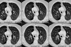
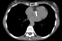
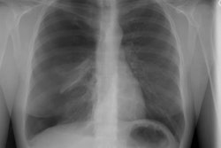
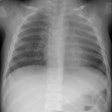
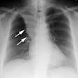
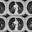
In many cases, the plain films will be unrevealing- radiographs provide significant information that alters treatment in 5% or fewer patients with acute asthma [3]. In cases in which abnormalities are present, plain film findings include bronchial wall thickening (48-71% of patients) [5], generalized hyperaeration (up to 24% of patients) [5], areas of over-inflation coupled with areas of atelectasis (due to mucous plugs obstructing the bronchi), atelectasis alone, and thickening of the interstitial tissues of the lungs. Mild prominence of the hilar vessels can be seen due to a transient pulmonary hypertension. Pneumomediastinum is occasionally noted as a complication, but pneumothorax is rare. Asthma may also lead to the chronic presence of patchy interstitial densities.HRCT findings include bronchial wall thickening (a subjective finding in up to 44-92% of asthmatic patients, but only 4-19% of controls) and evidence of air trapping/hypoxic vasoconstriction which produces a pattern of mosaic attenuation [2,5]. Mild bronchial dilatation can be seen in up to 77% of patients, but this finding may be related to hypoxic vasoconstriction of the adjacent pulmonary artery [5].
REFERENCES:
(1) Radiology 1997; 203:361-367
(2) Radiol Clin North Am 1994; Jul 32(4):745-757
(4) J Comput Assist Tomogr 1997; Kim Y, et al. The spectrum of eosinophilic lung disease: Radiologic findings. 21 (6): 920-930
(5) Radiol Clin North Am 1998; Lynch DA. Imaging of asthma and allergic bronchopulmonary mycosis. 36 (1): 129-142
(6) Society of Thoracic Radiology Course Syllabus 1999; Lynch DA. Imaging of asthma and its complications. 31-41 (No abstract available)
(7) Silva CI, et al. Asthma and associated conditions: high-resolution CT and pathologic findings. 183: 817-824

