AuntMinnie
  • Europe
  • Clinical News
  • Informatics
  • Industry News
  • Practice Management
  • Education
  • Subspecialties
  • More
  • Sign In
  • Board Review
  • CME
  • Careers
  • Cases
  • Conferences
  • Videos
  • Webinars
  • Podcast Network
  • Advertising
  • Buyer's Guide
  • Vendors
  • Minnies
Clinical News
  • CT
  • Digital X-Ray
  • Interventional
  • Molecular Imaging
  • MRI
  • Radiation Oncology/Therapy
  • Ultrasound
  • Womens Imaging
Imaging Informatics
  • Advanced Visualization
  • Enterprise Imaging
  • AI
  • Cybersecurity
Industry News
  • Installations
  • Product News
  • Regulatory
  • M&A
  • Market Analysis
Practice Management
  • Administration
  • Associations
  • Careers
  • Equity & Inclusion
  • Legislation
  • Medicolegal
  • Patient Safety
  • Radiologic Technologist
  • Service
  • Salary Scan
Resources
  • Board Review
  • CME
  • Careers
  • Cases
  • Conferences
  • Videos
  • Webinars
  • Podcast Network
  • Advertising
  • Buyer's Guide
  • Vendors
  • Minnies
User Tools
  • Sign In
More
  • About Us
  • Advertising
  • Help
Follow AuntMinnie
Facebook iconTwitter X icon LinkedIn iconInstagram icon
  • Europe
  • Clinical News
    • CT
    • Digital X-Ray
    • Interventional
    • Molecular Imaging
    • MRI
    • Radiation Oncology/Therapy
    • Ultrasound
    • Womens Imaging
  • Informatics
    • Advanced Visualization
    • Enterprise Imaging
    • AI
    • Cybersecurity
  • Industry News
    • Installations
    • Product News
    • Regulatory
    • M&A
    • Market Analysis
  • Practice Management
    • Administration
    • Associations
    • Careers
    • Equity & Inclusion
    • Legislation
    • Medicolegal
    • Patient Safety
    • Radiologic Technologist
    • Service
    • Salary Scan
  • Education
    • Medical Students
    • Residents/Fellows
  • Subspecialties
    • Breast Imaging
    • CV
    • Chest
    • Emergency
    • GI
    • GU
    • Head & Neck
    • Interventional
    • Physics
    • MSK
    • Neuro
    • Nuclear
    • Pediatric
    • Radiation Oncology
  • More
  • Board Review
  • CME
  • Careers
  • Cases
  • Conferences
  • Videos
  • Webinars
  • Podcast Network
  • Advertising
  • Buyer's Guide
  • Vendors
  • Minnies
  • Sign In
Follow AuntMinnie
Facebook iconTwitter X icon LinkedIn iconInstagram icon

Brian Casey

[email protected]
Musculoskeletal Radiology
Ultrasound performs well for wrist pain, meniscal tears
February 4, 2008
Digital X-Ray
Buyer's Guide Update: Angio systems go digital, get therapeutic
February 3, 2008
Digital X-Ray
Dark-field x-ray measures scatter radiation for better image quality
A new technique for producing images could offer a way to create higher-quality images using conventional digital radiography equipment, according to Swiss researchers. The technique, called dark-field x-ray, is based on radiation scattered from a target rather than the traditional method of using radiation absorbed by the target. The researchers discussed their implementation of dark-field imaging in a paper published in Nature Materials.
February 3, 2008
2008 01 29 16 37 07 706
Molecular Imaging
PET/CT shows value in head and neck tumors
January 30, 2008
Digital X-Ray
AuntMinnie.com Digital X-Ray Insider
January 29, 2008
Womens Imaging
The return of SalaryScan; update on DMIST results
January 28, 2008
IS
Data mining your reports; more on NSF and gadolinium
January 23, 2008
AI
CAD boosts accuracy without increasing recalls
January 21, 2008
Cardiac Imaging
The clinical argument for coronary CTA
January 16, 2008
Practice Management
Maryland law out-Starks Stark; last chance to take personality survey
January 14, 2008
Previous Page
Page 85 of 190
Next Page
Top Stories
2026 02 12 Chest Ac2 Thumbnail
Digital X-Ray
Chest x-rays reveal atherosclerosis in patients undergoing amputations
Lower limb amputation is an increasingly common major complication of advanced peripheral arterial disease and diabetes mellitus.
Cardiac Mri Adobe Samunella
MRI
3D MRI technique helps plan treatment for pediatric heart conditions
Images of a 39-year-old who underwent screening mammography. Right craniocaudal (left) and mediolateral oblique (middle) views from preoperative mammography show suspicious grouped fine pleomorphic calcifications (arrows). After biopsy showed ductal carcinoma of situ (DCIS), patient underwent breast-conserving surgery, yielding final diagnosis of ductal carcinoma in situ (intermediate grade, hormone receptor positive). (Right) Screenshot of an output of a commercial AI tool applied to preoperative mammography. AI detected right breast lesion with score of 95% on both views (concentric colored circles). Present study’s reviewing radiologist deemed AI marking to correspond with site of DCIS.
Womens Imaging
Could AI scoring help with managing DCIS?
Axial contrast-enhanced CT scan in a 28-year-old premenopausal woman shows an incidentally detected solid-appearing left ovarian lesion (solid arrow) anterior to the uterus (U), associated with trace ascites in the right adnexa and cul de sac (dashed arrows). At pathologic analysis, this was shown to be ovarian dysgerminoma. Two readers misdiagnosed this lesion, one as a leiomyoma and one as an ovarian fibroma.
CT
Clinicians, beware: CT diagnostic accuracy varies by adnexal lesion type
Erin Stephens
Practice Management
Radiology coding update for 2026
More in Home
Brain MRI tracks risk of stage IV breast cancer metastases
By Kate Madden Yee
Brain MRI shows promise for surveilling women at risk for asymptomatic stage IV breast cancer metastases by tracking molecular subtypes of the disease.
January 24, 2025
Brain Mri
Experts call for CPT codes for imaging AI reimbursement
By Amerigo Allegretto
Imaging AI tools and algorithms continue to be rapidly developed, but experts say reimbursement through CPT codes is needed.
January 24, 2025
Insurance Computer 400
Is herpes infection protective in early Alzheimer’s disease patients?
By Will Morton
In a recent PET study with “unexpected” results, patients infected with herpes had fewer signs of brain deposits associated with Alzheimer’s disease than uninfected patients.
January 24, 2025
Amyloid
Triple-negative breast cancers impact female populations differently
By Amerigo Allegretto
Despite overall breast cancer rate increases, women of varying race and ethnicity experience disproportionately higher increases.
January 24, 2025
Breast Cancer Cell2 Social
What brain mechanisms drive depression in older adults?
By Will Morton
PET has revealed that mechanisms other than amyloid brain deposits may underlie depressive symptoms in people later in life.
January 23, 2025
Ad
MR spectroscopy reveals neurochemical patterns linked to COVID-19
By Kate Madden Yee
Proton MR spectroscopy (MRS) reveals "remarkable" neurochemical patterns associated with COVID-19-induced anosmia, or loss of smell.
January 23, 2025
Mri Brain
LI-RADS ultrasound surveillance version 2024 improves HCC detection
By Amerigo Allegretto
The 2024 version of the LI-RADS Ultrasound Surveillance system improves detection of hepatocellular carcinoma (HCC) over earlier iterations.
January 22, 2025
(A,B) Images from an initial ultrasound examination showed no identifiable lesions and received a consensus ultrasound category of US-1. Nearly the entire liver is visualized, with minimal shadowing (“s”, A and B), and the examination received consensus result of VIS-A. The AFP level was 7.1 ng/mL on assessment six months before the ultrasound, 9.1 ng/mL on assessment three months before examination, and 14.2 ng/mL on assessment on exam day. The AFP result was thus assessed as positive based on a progressive increase on two consecutive tests. The atient was considered to have negative surveillance result according to LI-RADS Ultrasound Surveillance v2017, but positive surveillance result according to LI-RADS Ultrasound Surveillance v2024. (C-D) Axial arterial-phase (C) and portal-venous phase (D) T1-weighted images from subsequent gadoxetic acid-enhanced MRI show a 2-cm arterial-phase hyperenhancing observation (arrow, C) with portal-venous washout (arrow, D) in segment 7. This observation was classified on MRI as LR-5, thus meeting the present study’s reference standard for diagnosis of HCC. The mass is located in the periphery of the liver, an anatomic area where ultrasound visualization can be challenging due to shadowing.
Deep learning helps find, segment lung tumors on CT imaging
By Kate Madden Yee
Using a deep-learning model with lung CT imaging shows promise for identifying and segmenting tumors, researchers have reported.
January 22, 2025
Lung Ct Images
EVT promising in patients with extracranial ICA occlusions
By Will Morton
Endovascular thrombectomy (EVT) and best medical treatment are both effective and safe options for patients with acute extracranial internal carotid artery occlusions.
January 21, 2025
Images in a 74-year-old male patient who presented at the hospital with acute symptoms of a right hemispheric ischemic stroke and a National Institutes of Health Stroke Scale (NIHSS) score of 24. (A) Nonenhanced CT image shows no signs of early infarction, whereas (B) CT perfusion image shows a large perfusion deficit in the territory of the middle cerebral artery and anterior cerebral artery caused by an extracranial occlusion in the right internal carotid artery. (C) Digital subtraction angiography shows the occlusion in the C1 segment. The extracranial occlusion was passed, reperfused, and subsequently stented (D, E). The final contrast injection did not reveal any intracranial occlusions within the territory of the internal carotid artery (F). At the 24-hour follow-up, there was no infarction on (G) the nonenhanced CT image, and the NIHSS score had decreased to 3 points. Image courtesy of RSNA.
CEUS resolves indeterminate CT, MRI liver observations
By Amerigo Allegretto
Contrast-enhanced ultrasound (CEUS) can resolve some high-risk indeterminate liver observations initially seen on CT or MRI.
January 21, 2025
Images depict a 49-year-old female participant with cryptogenic cirrhosis who presented with a 3.2-cm liver lesion. This biopsy-confirmed hepatocellular carcinoma (HCC) with an initial categorization of Liver Imaging Reporting and Data System (LI-RADS) category LR-4 (probably HCC) at CT was upgraded to LR-5 (definitely HCC) at contrast-enhanced ultrasound (CEUS). (A) Precontrast CT images in the liver show vague hypoattenuation in the medial left hepatic lobe (arrows). (B) Arterial phase images demonstrate no definite arterial phase hyperenhancement (arrows). (C) Washout and an enhancing capsule (arrows) on a late-phase image resulted in an LR-4 categorization. (D) B-mode ultrasound shows a round hypoechoic nodule (arrows). (E) An early-phase CEUS image shows clear arterial-phase hyperenhancement (arrows); (F) No washout at one minute (arrows), and (G) late, mild washout at four minutes (arrows) resulted in a CEUS LR-5 categorization.
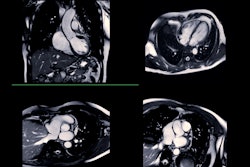
PET/CT can rule out CAV in heart transplant patients
By Will Morton
Cardiac PET/CT scans can effectively rule out moderate to severe cardiac allograft vasculopathy (CAV) in heart transplant patients.
January 17, 2025
Ctca
Machine learning can predict immunotherapy response in NSCLC patients
By Kate Madden Yee
Machine learning can help clinicians predict immunotherapy response in patients with inoperable non-small cell lung cancer (NSCLC).
January 17, 2025
Lung Ct Images
Previous Page
Page 85 of 202
Next Page
AuntMinnie
Topics
  • Clinical News
  • Imaging Informatics
  • Industry News
  • Practice Management
  • Radiology Education
  • Subspecialties
Resources
  • Board Review
  • CME
  • Careers
  • Cases
  • Conferences
  • Videos
  • Webinars
  • Podcast Network
  • Advertising
  • Buyer's Guide
  • Vendors
  • Minnies
More
  • About Us
  • Advertising
  • Help
Follow AuntMinnie
Facebook iconTwitter X icon LinkedIn iconInstagram icon
  1. Privacy Policy
  2. Privacy Settings
  3. Terms & Conditions
  4. Contact Us
  5. Site Map
  6. Home
© 2026 Science and Medicine Group. All rights reserved.