Contrast-enhanced ultrasound (CEUS) can resolve some high-risk indeterminate liver observations initially seen on CT or MRI, suggest findings published January 21 in Radiology.
Researchers led by Andrej Lyshchik, MD, PhD, from Thomas Jefferson University Hospital in Philadelphia, PA found that CEUS had higher clinical impact for high-risk CT and MRI liver observations measuring at least 20 mm.
“These findings highlight the significant clinical value of CEUS in the management of high-risk liver lesions with indeterminate CT/MRI LI-RADS categorization,” Lyshchik and colleagues wrote.
Radiologists continue to explore the potential of CEUS in liver imaging, particularly in detecting hepatocellular carcinoma (HCC). The modality is used to categorize liver nodules depicted at screening ultrasound or diagnostic CT or MRI. The researchers highlighted several advantages of using CEUS in this area. These include its ability to allow continuous imaging and precise correlation of arterial phase hyperenhancement with the time of occurrence, high spatial and temporal resolution, and high sensitivity to contrast agents.
Lyshchik and co-authors studied the clinical impact of CEUS in patients with high-risk indeterminate liver observations categorized as LR-4 at CT (probably HCC) or LR-M at MRI (probably or definitely malignant but not HCC specific).
The study is a secondary analysis of a prospective international multicenter validation study for CEUS LI-RADS performed between 2018 and 2021.
Radiologists performed CEUS within four weeks of CT or MRI. The researchers used data from tissue histologic analysis and follow-up CT or MRI as reference standards. They also evaluated the clinical effect of CEUS for HCC analysis in liver observations 10 mm or larger categorized as CT/MRI LR-4 and LR-M.
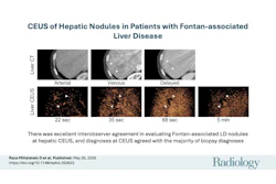
 Images depict a 49-year-old female participant with cryptogenic cirrhosis who presented with a 3.2-cm liver lesion. This biopsy-confirmed hepatocellular carcinoma (HCC) with an initial categorization of Liver Imaging Reporting and Data System (LI-RADS) category LR-4 (probably HCC) at CT was upgraded to LR-5 (definitely HCC) at contrast-enhanced ultrasound (CEUS). (A) Precontrast CT images in the liver show vague hypoattenuation in the medial left hepatic lobe (arrows). (B) Arterial phase images demonstrate no definite arterial phase hyperenhancement (arrows). (C) Washout and an enhancing capsule (arrows) on a late-phase image resulted in an LR-4 categorization. (D) B-mode ultrasound shows a round hypoechoic nodule (arrows). (E) An early-phase CEUS image shows clear arterial-phase hyperenhancement (arrows); (F) No washout at one minute (arrows), and (G) late, mild washout at four minutes (arrows) resulted in a CEUS LR-5 categorization.RSNA
The study included data collected from 109 patients with an average age of 64.3 years. This also included 113 observations that were 10 mm or larger and were categorized as LR-4 on CT (n = 60) and LR-M on MRI (n = 53).
CEUS resulted in management recommendation changes in 38 of these observations (33.6%). Among these, 36 were correct.
The team also reported that 34 of the 113 observations on CT/MRI were categorized on CEUS as LI-RADS 5, meaning definite HCC. This made biopsy unnecessary. Of these LI-RADS 5 exams, CEUS correctly categorized 32 of them.
Finally, the researchers highlighted that CEUS had more clinical impact for liver observations 20 mm or larger (n = 68). Here, CEUS helped appropriately categorize both LR-5 and LR-M lesions as HCC and non-HCC malignancies. This resulted in management recommendation changes in 40% of observations (n = 27) with 100% accuracy.
“CEUS LI-RADS showed the ability to help conclusively diagnose HCC, alleviating the need for tissue sampling, or correctly referring to observations for biopsy,” the study authors wrote.
They added that the findings support the clinical value of CEUS in managing high-risk liver lesions with indeterminate categorization on CT or MRI. The authors called for future studies to evaluate interreader agreement for CEUS LI-RADS and “possibly” intermodality with CT/MRI LI-RADS.
The full study can be accessed here.