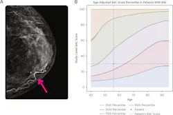
Legislation introduced in the state of Maryland would require women to be informed if their mammogram results show breast arterial calcification (BAC).
House Bill (HB) 1364 includes provisions that model breast density notification by letter after a mammography exam. It was introduced on February 13.
"The bill would require notification to women in their mammogram results letter if breast arterial calcification, known as BAC, is present in their mammogram," Joseline Pena-Melnyk, speaker of the Maryland House of Delegates, told the House Health Committee on February 24. "Although BAC frequently appears on routine mammograms, there is currently no standardized protocol for reporting this information to a patient. Even when the presence is documented, reporting practices lack uniformity. This bill will provide that needed uniformity."
The language included in HB 1364 is intended to raise nonalarming awareness that BAC is a common finding and that it may be an indicator of increased risk of cardiovascular disease. As written currently, the bill asks recipients to discuss the finding with their physician to assess cardiovascular risks and determine if additional testing is appropriate.
If passed, the act would go into effect on October 1, 2026.

![A normal mammogram confirmed by three-year radiologic follow-up illustrates reader-marked regions of interest (ROIs) during (A) unaided (round 1) and (B) artificial intelligence (AI)–assisted (round 2) reading. Each colored dot represents an ROI for recall by a human reader. Readers could mark more than one ROI per case, represented by multiple dots of the same color. During AI-assisted reading, the AI system displayed three visible prompts: two with suspicion of malignancy scores of 35% (left mediolateral oblique [L MLO] and craniocaudal [L CC]) and one with a suspicion of malignancy score of 10% (right craniocaudal [R CC]), shown as polygonal overlays. Without AI, six of 10 readers (60%) marked a false-positive ROI. With AI assistance, this fell to two of 10 (20%). R MLO = right mediolateral oblique.](https://img.auntminnie.com/mindful/smg/workspaces/default/uploads/2026/07/2026-07-14-radiology-mammogram-ai-auto-bias.H0bYO8QlWs.jpg?auto=format%2Ccompress&fit=crop&h=100&q=70&w=100)


![A normal mammogram confirmed by three-year radiologic follow-up illustrates reader-marked regions of interest (ROIs) during (A) unaided (round 1) and (B) artificial intelligence (AI)–assisted (round 2) reading. Each colored dot represents an ROI for recall by a human reader. Readers could mark more than one ROI per case, represented by multiple dots of the same color. During AI-assisted reading, the AI system displayed three visible prompts: two with suspicion of malignancy scores of 35% (left mediolateral oblique [L MLO] and craniocaudal [L CC]) and one with a suspicion of malignancy score of 10% (right craniocaudal [R CC]), shown as polygonal overlays. Without AI, six of 10 readers (60%) marked a false-positive ROI. With AI assistance, this fell to two of 10 (20%). R MLO = right mediolateral oblique.](https://img.auntminnie.com/mindful/smg/workspaces/default/uploads/2026/07/2026-07-14-radiology-mammogram-ai-auto-bias.H0bYO8QlWs.jpg?auto=format%2Ccompress&fit=crop&h=112&q=70&w=112)