The American College of Radiology (ACR), Breast Cancer Research Foundation (BCRF), and GE HealthCare are enrolling the first patients in the Contrast-Enhanced Mammography Imaging Screening Trial (CMIST).
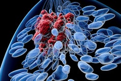
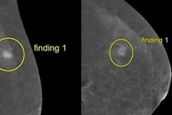
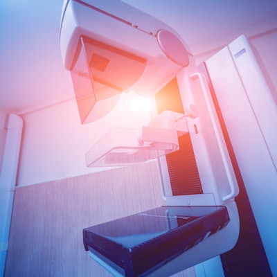
The study aims to find out whether contrast-enhanced mammography (CEM) helps with breast cancer detection and reduction false-positive exams in women with dense breasts. Standard mammography struggles with imaging dense breasts, meaning breast density is a risk factor in breast cancer development.
The U.S. Food and Drug Administration (FDA) recently announced a change in the Mammography Quality Standards Act (MQSA) that will require patient notification about their breast density.
CEM adds iodinated contrast media to mammography, highlighting blood flow patterns in breast tissue. The researchers aim to enroll a total of 2,032 women with dense breasts in CMIST, comparing CEM to 3D mammography.
The group also indicated that 15 sites in the U.S. and Canada are planned to start recruiting patients, with Carolina Breast Imaging Specialists in Greenville, North Carolina being the first. The team said it plans to publish the first results in 2025.

![A normal mammogram confirmed by three-year radiologic follow-up illustrates reader-marked regions of interest (ROIs) during (A) unaided (round 1) and (B) artificial intelligence (AI)–assisted (round 2) reading. Each colored dot represents an ROI for recall by a human reader. Readers could mark more than one ROI per case, represented by multiple dots of the same color. During AI-assisted reading, the AI system displayed three visible prompts: two with suspicion of malignancy scores of 35% (left mediolateral oblique [L MLO] and craniocaudal [L CC]) and one with a suspicion of malignancy score of 10% (right craniocaudal [R CC]), shown as polygonal overlays. Without AI, six of 10 readers (60%) marked a false-positive ROI. With AI assistance, this fell to two of 10 (20%). R MLO = right mediolateral oblique.](https://img.auntminnie.com/mindful/smg/workspaces/default/uploads/2026/07/2026-07-14-radiology-mammogram-ai-auto-bias.H0bYO8QlWs.jpg?auto=format%2Ccompress&fit=crop&h=100&q=70&w=100)


![A normal mammogram confirmed by three-year radiologic follow-up illustrates reader-marked regions of interest (ROIs) during (A) unaided (round 1) and (B) artificial intelligence (AI)–assisted (round 2) reading. Each colored dot represents an ROI for recall by a human reader. Readers could mark more than one ROI per case, represented by multiple dots of the same color. During AI-assisted reading, the AI system displayed three visible prompts: two with suspicion of malignancy scores of 35% (left mediolateral oblique [L MLO] and craniocaudal [L CC]) and one with a suspicion of malignancy score of 10% (right craniocaudal [R CC]), shown as polygonal overlays. Without AI, six of 10 readers (60%) marked a false-positive ROI. With AI assistance, this fell to two of 10 (20%). R MLO = right mediolateral oblique.](https://img.auntminnie.com/mindful/smg/workspaces/default/uploads/2026/07/2026-07-14-radiology-mammogram-ai-auto-bias.H0bYO8QlWs.jpg?auto=format%2Ccompress&fit=crop&h=112&q=70&w=112)