Monday, December 2 | 3:20 p.m.-3:30 p.m. | SSE02-03 | Room E451B
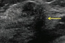
Do women with dense breast tissue who have a normal digital breast tomosynthesis (DBT) exam need supplemental whole-breast ultrasound (WBUS) imaging? Perhaps not, according to research to be presented Monday afternoon.Presenter Dr. Liane Philpotts of Yale University and colleagues conducted a study that explored the performance outcomes of supplemental handheld WBUS screening in women with dense tissue after a normal tomosynthesis screening mammogram. The study included 5,742 WBUS exams acquired between October 2017 and September 2018; the group tracked final BI-RADS assessments, lesion size and type, and outcome of BI-RADS 3 (probably benign) and 4 (suspicious) cases.
The majority of the WBUS exams' final BI-RADS assessments were 1 or 2 (97.2%); 2.4% were BI-RADS 3 and 0.4% were BI-RADS 4. Two cancer cases were identified, for a detection rate of 0.7 per 1,000 exams.
The findings raise the question of whether supplemental ultrasound after a normal DBT exam in women with dense tissue is needed, Philpotts and colleagues concluded.
"After normal tomosynthesis mammograms, the majority of WBUS cases were found to be normal, with only a small proportion of cases requiring follow-up or biopsy," they wrote in their abstract.